May-Thurner syndrome (MTS) is an underdiagnosed vascular condition that affects mcv blood test results meaning and normal range flow in the pelvis, primarily causing compression of the left common iliac vein by the right common iliac artery1. This compression can lead to serious complications such as deep vein thrombosis (DVT) and pulmonary embolism (PE), especially in young to middle-aged adults2. While many people with MTS remain asymptomatic, those affected often experience symptoms related to impaired venous return and venous obstruction in the left leg3.
Signs and Symptoms
May-Thurner syndrome presents with a range of symptoms that vary from none at all to severe venous stasis and thrombosis4. Most patients experience symptoms on the left lower extremity, reflecting the typical anatomical site of venous compression5. Common clinical features include unilateral leg swelling, pain, and visible varicose veins6. Patients often describe a sensation of heaviness or fullness in the affected leg, sometimes accompanied by dull, aching, or throbbing pain54. Skin changes such as redness, hyperpigmentation, or purple spots may also be observed5.
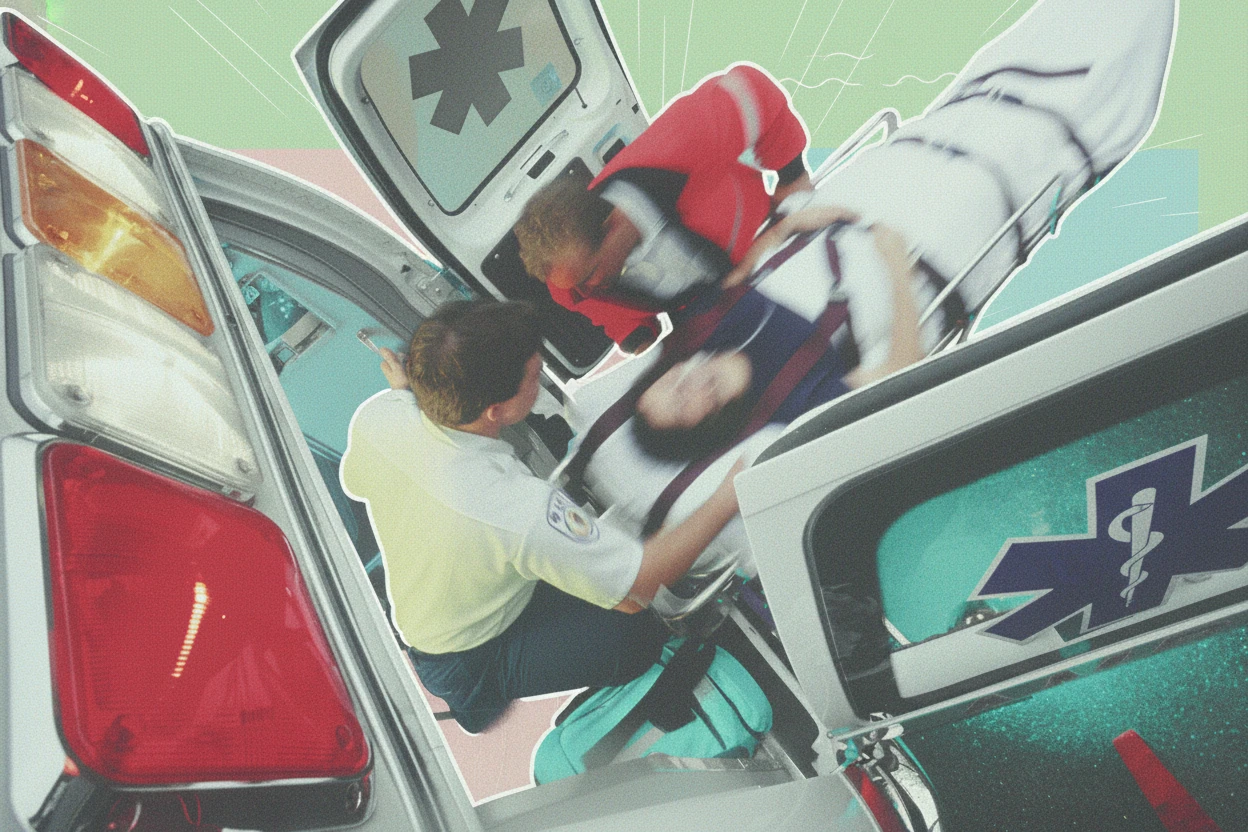
Chronic cases of MTS can lead to venous claudication—pain during walking due to poor venous blood flow—and venous stasis ulcers, which are slow-healing sores resulting from prolonged venous insufficiency78. In some instances, MTS remains silent until a deep vein thrombosis develops, often presenting as sudden leg swelling and pain910.
Causes and Risk Factors
May-Thurner syndrome is caused by the anatomical compression of the left common iliac vein by the overlying right common iliac artery as it crosses the pelvis5. This compression narrows the vein (venous stenosis), impairs blood flow, and can injure the venous endothelium, promoting the formation of fibrous bands or spurs inside the vein1. These changes increase the risk of venous obstruction and thrombosis.
Risk Factors
- Female sex: MTS is more common in females, with a female-to-male ratio of approximately 5:1511.
- Age: Most patients present between 20 and 50 years of age511.
- Pregnancy: The increased pelvic pressure and vascular changes during pregnancy can contribute to venous compression511.
- Oral contraceptive use: Hormonal factors increase the risk of thrombosis in MTS511.
- Dehydration: Reduced blood volume can increase clotting risk in affected individuals511.
- Prolonged immobility and obesity: These conditions promote venous stasis and thrombosis risk1213.
- Anatomical variations: Congenital or acquired pelvic vascular variants may predispose to MTS511.
The left iliac vein is responsible for venous return from the lower extremities, and its compression leads to impaired blood flow and venous hypertension5. This mechanical obstruction is a significant risk factor for left-sided deep vein thrombosis5.
Diagnostic Process
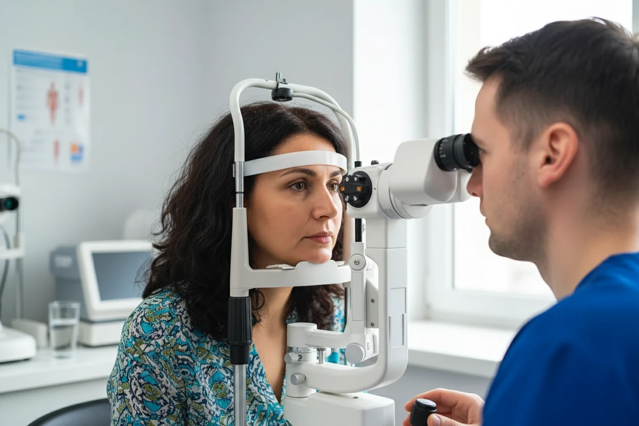
Diagnosing May-Thurner syndrome involves clinical evaluation combined with imaging studies to confirm venous compression and assess for thrombosis511. Physical examination may reveal unilateral leg edema, varicosities, and skin changes such as redness or hyperpigmentation5. Signs of acute deep vein thrombosis include tenderness, warmth, and erythema of the affected limb5.
Duplex ultrasound is the first-line imaging modality used to evaluate venous flow, detect stenosis, and identify thrombus formation511. However, ultrasound may have limitations in visualizing pelvic veins. Therefore, CT venography is often employed for detailed imaging of the iliac veins, providing high sensitivity and specificity to detect venous compression, stenosis, collateral vessels, and DVT5116. Magnetic resonance venography (MRV) is an alternative imaging option that offers detailed visualization without radiation exposure511.
Elevated D-dimer levels in blood tests can indicate recent or ongoing thrombosis, supporting the diagnosis of DVT in MTS patients511.
Stages of May-Thurner Syndrome
May-Thurner syndrome progresses through three clinical stages121314:
| Stage | Description | Clinical Features |
|---|---|---|
| I | Asymptomatic left iliac vein compression | No symptoms, incidental finding |
| II | Formation of venous spurs and stenosis | Venous insufficiency, edema, varicosities |
| III | Left lower extremity deep vein thrombosis (DVT) | Acute leg swelling, pain, risk of PE |
| Sources:121314 | ||
This progression reflects increasing severity from mechanical compression without symptoms to significant venous obstruction and thrombosis115.
May-Thurner syndrome often remains undiagnosed until a patient develops a deep vein thrombosis, highlighting the importance of awareness and early imaging in patients with unexplained unilateral leg swelling1016.
Treatment Options
Treatment of May-Thurner syndrome aims to restore normal venous blood flow, relieve symptoms, and reduce the risk of thrombosis and its complications5116. Management depends on the presence or absence of deep vein thrombosis and symptom severity.
Medical Procedures
Endovascular treatment is the preferred approach for symptomatic MTS5116. This typically involves angioplasty, where a balloon is used to dilate the compressed iliac vein, followed by stent placement to maintain patency and prevent re-compression5116. These minimally invasive procedures have high success rates and low complication rates compared to open surgery17.
Surgical bypass or vein repositioning is reserved for patients who do not respond to endovascular therapy or have complex anatomical issues5116. Surgical options are less common due to advances in endovascular techniques1213.
Medications
For patients with acute deep vein thrombosis, anticoagulation therapy is essential to prevent clot extension and new thrombus formation5116. Thrombolytic therapy (clot-busting drugs) may be used in selected cases to rapidly dissolve large clots5116. However, anticoagulation alone is insufficient in MTS without correction of the underlying venous compression1.
Compression Socks
Compression stockings are often recommended to help manage symptoms by improving venous return and reducing leg swelling5116. They are especially useful in chronic venous insufficiency and post-thrombotic syndrome but do not address the mechanical cause of MTS511.
May-Thurner Syndrome is increasingly recognized as a cause of chronic venous insufficiency and a precipitating factor for venous thromboembolism. Despite controversy about the exact definition of the pathology, this review confirms endovascular treatment to be safe and effective therapy for acute venous thrombosis or chronic compression17.
Prevention Strategies
Preventing complications of May-Thurner syndrome focuses on reducing risk factors for venous thrombosis and promoting healthy venous circulation5116. Lifestyle modifications include:
- Regular exercise to enhance venous return and reduce blood pooling5116.
- Avoiding prolonged immobility, especially during travel or after surgery5116.
- Maintaining adequate hydration to support normal blood viscosity5116.
- Managing obesity to decrease venous pressure and thrombosis risk5116.
- Smoking cessation to improve vascular health and reduce clotting risk5116.
- Following a balanced diet to support cardiovascular and venous function5116.
These measures help minimize the risk of DVT and other venous complications associated with MTS.
Associated Conditions
May-Thurner syndrome is linked to several venous disorders due to impaired blood flow and venous stasis5116. The most common associated conditions include:
- Deep vein thrombosis (DVT), particularly on the left side, due to venous obstruction5116.
- Chronic venous insufficiency (CVI), characterized by venous pooling and leg swelling5116.
- Pulmonary embolism (PE), a life-threatening complication when a clot dislodges and travels to the lungs5116.
- Post-thrombotic syndrome (PTS), a chronic condition with pain, swelling, and skin changes following DVT5116.
Early diagnosis and treatment of MTS reduce the risk of these complications and improve long-term outcomes1.
Daily Management
Living with May-Thurner syndrome involves ongoing care to manage symptoms and prevent complications7. Patients are encouraged to:
- Use compression stockings regularly to reduce leg swelling and discomfort5116.
- Elevate the legs when resting to improve venous return5116.
- Engage in regular physical activity tailored to their ability5116.
- Maintain hydration and a healthy weight5116.
- Attend regular follow-up appointments for monitoring and imaging as needed5116.
These strategies support venous health and enhance quality of life for individuals with MTS.
Amy Bonner’s story illustrates successful management of MTS with catheter-directed clot removal and stenting, leading to long-term symptom relief and improved quality of life18.
Common Questions
Most patients with May-Thurner syndrome have a good prognosis when appropriately treated121314. With timely intervention and lifestyle modifications, individuals can expect a normal lifespan and quality of life5116. Adherence to treatment plans, including anticoagulation when indicated and use of compression therapy, is key to preventing recurrent thrombosis and chronic venous complications5116.
Regular exercise, a balanced diet, and smoking cessation are recommended to support vascular health5116. Compression stockings and leg elevation help manage symptoms and reduce edema5116. Ongoing medical follow-up is important to monitor for disease progression or complications5116.