Breast implants are common, with nearly 300,000 women undergoing augmentation annually in the US. While implants do not increase breast cancer risk, they can affect how breast cancer is detected during screening, making awareness and proper imaging techniques essential12. Understanding how implants influence mammography and other screening methods helps ensure early detection and effective management3.
Understanding Your Implants' Feel
After breast augmentation or reconstruction, it is important for patients to become familiar with how their breasts feel to detect any changes. Implants can sometimes be mistaken for lumps during self-exams, causing confusion2. Saline implants may feel like a water balloon because of their fluid content and can bulge under pressure2. Patients should learn to distinguish implant from natural breast tissue, ideally with guidance from their surgeon2.
- Knowing the normal feel of your implants helps identify new lumps or changes2.
- Smaller implants may make it easier to detect breast lumps during self or clinical exams2.
- Any new lump should be promptly evaluated by a healthcare professional and not dismissed as implant-related without assessment2.
- Regular self-exams remain important even with implants3.
Implants Can Reduce Detection Accuracy
Breast implants can obscure mammographic images, reducing the ability to detect cancer deaths2. The location of cancer relative to the implant affects its visibility on mammograms, making detection more challenging2. Mammography sensitivity in women with implants is lower—about 77.8%—compared to roughly 90.7% in women without implants41. Despite this, breast cancer prognosis does not differ significantly between women with or without implants2.
- Implants block X-rays from fully penetrating breast tissue, especially if placed in front of chest muscles3.
- Cancers may be missed or detected later due to reduced mammographic sensitivity1.
- Breast cancers detected in women with implants tend to be smaller at diagnosis, especially when found by self-exam or clinical exam1.
- The type of implant (silicone vs. saline) or implant placement (subpectoral vs. subglandular) does not significantly affect cancer detection, though saline implants may allow slightly better mammographic visualization15.
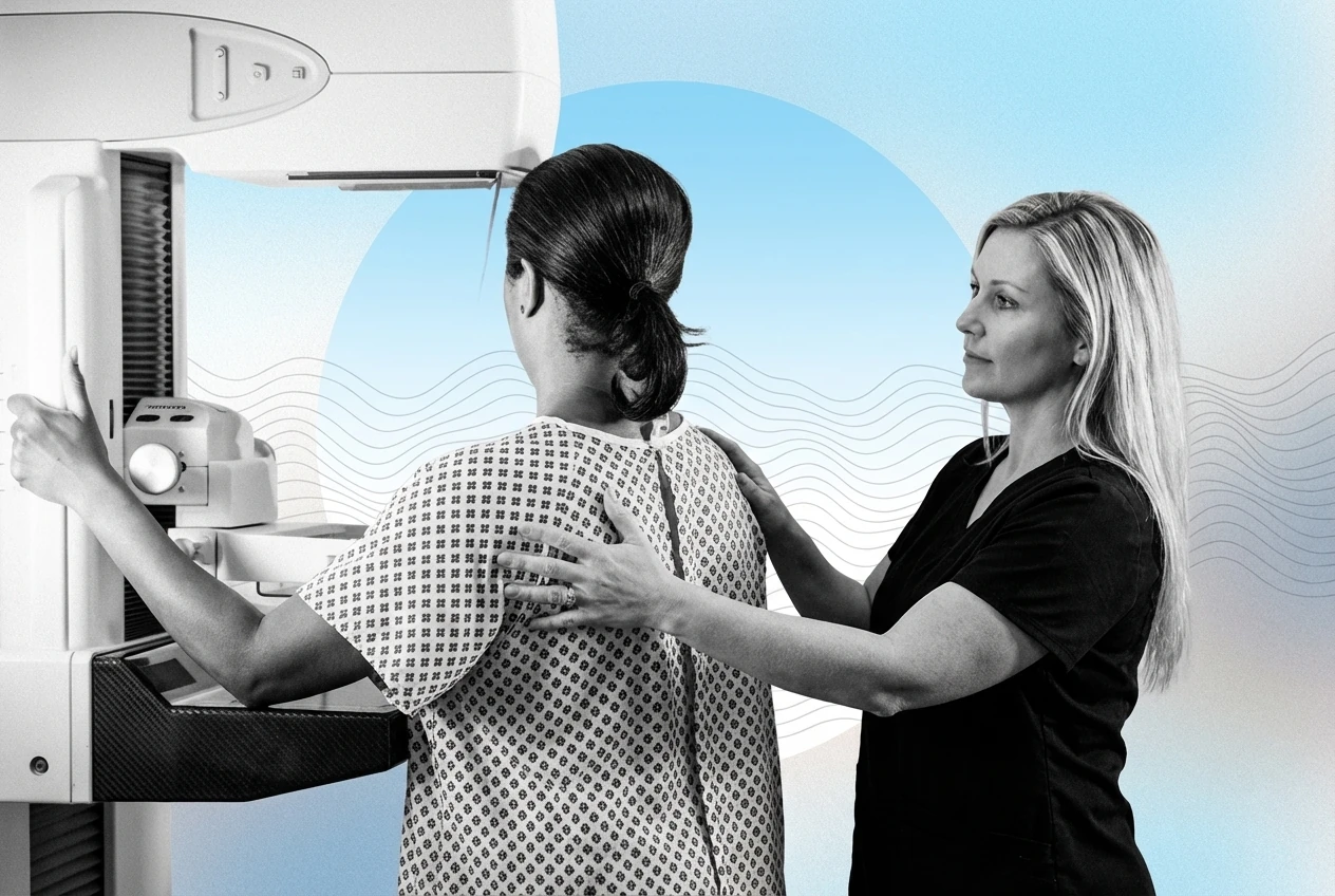
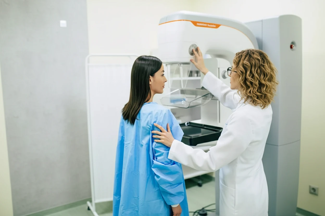
Notify Your Mammography Technicians
Informing the mammography team about your implants before the procedure is critical. This allows technologists to adjust positioning and compression techniques to optimize imaging while minimizing risk23.
- Tell the scheduler and technologist you have implants when making and arriving for your appointment23.
- Experienced technologists know how to position patients and adjust compression to avoid implant damage23.
- Proper communication helps ensure the best possible images of breast tissue around implants2.
- Facilities with experience in imaging patients with implants provide safer and more effective screening3.
Additional Imaging May Be Required
Women with breast implants often need extra mammographic views to visualize breast tissue hidden behind implants. These additional images, called implant displacement views, involve pushing the implant back against the chest wall and pulling the breast tissue forward23.
- Standard mammography includes two views per breast; implant displacement adds two more views per breast23.
- Implant displacement views use minimal compression to avoid implant rupture while stabilizing breast tissue2.
- Proper patient positioning is essential to maximize breast tissue visualization2.
- Technologists trained in implant imaging techniques reduce the risk of implant damage2.
“Women with breast implants typically require additional mammographic views to improve cancer detection, but these extra images are safe and carefully performed.”67
Mammography Implant Rupture Risk Is Low
Concerns about implant rupture during mammography are common but largely unfounded. The Food and Drug Administration (FDA) reports very few cases of implant rupture linked to mammography compression23.
- Implant rupture during mammography is extremely rare28.
- Most ruptures occur in implants already weakened before the mammogram2.
- Mammography uses careful compression techniques to protect implants2.
- Routine mammography involves four images (two views per breast), with additional views for implants done with minimal pressure2.
- The benefits of mammography for early cancer detection far outweigh the minimal risk of rupture8.
Alternative Screening Methods Sometimes Needed
While mammography remains the primary screening tool, additional imaging may be necessary for women with implants, especially if mammograms are inconclusive or breast tissue is difficult to visualize.
Ultrasound
Ultrasound can detect masses obscured by implants on mammograms and is often used for women at high risk or with dense breast tissue29.
- Ultrasound is a supplemental screening tool, not a replacement for mammography9.
- It helps identify lesions hidden behind implants2.
- Typically reserved for women with dense breasts or elevated breast cancer risk9.
- Ultrasound cannot detect microcalcifications, which mammography can identify9.
Needle Biopsy
If imaging reveals suspicious areas, a needle biopsy may be performed to obtain tissue samples for diagnosis2.
- Needle biopsy near implants carries a small risk of implant rupture, though this is uncommon2.
- Biopsy provides definitive diagnosis when imaging is inconclusive2.
- Patients should discuss biopsy risks and benefits with their healthcare provider2.
Lymphoma Risk Association
Breast implants are not linked to an increased risk of breast cancer, but they are associated with a rare lymphoma called breast implant-associated anaplastic large cell lymphoma (BIA-ALCL)10211.
- BIA-ALCL is a T-cell lymphoma that develops in the scar tissue (capsule) around textured breast implants1011.
- Symptoms include breast asymmetry, pain, redness, swelling, or lumps near the implant1011.
- Treatment typically involves removal of the implant and capsule (capsulectomy)10.
- Advanced cases may require chemotherapy or radiation therapy10.
- As of 2023, the FDA has documented over 1,200 cases of BIA-ALCL with several fatalities1211.
- Regulatory actions have been taken to improve implant safety and require long-term studies12.
Removal Due to Complications
Certain complications related to breast cancer treatment or implant-associated conditions may necessitate implant removal.
- Implant removal decisions are individualized based on cancer subtype and treatment plans2.
- Radiation therapy increases the risk of capsular contracture, causing hardening and deformity of breast tissue around implants2.
- Severe capsular contracture can cause pain and visible breast shape changes2.
- Implants do not need to be removed solely to undergo mammographic screening2.
- Removing intact implants just for screening purposes is generally not recommended2.
Key Takeaways
- Breast implants can obscure mammographic images, reducing cancer detection sensitivity but do not increase breast cancer risk42.
- Patients should become familiar with their implants' feel to detect any new lumps or changes2.
- Informing mammography staff about implants ensures proper imaging techniques and reduces risk23.
- Additional mammographic views and supplemental imaging like ultrasound may be necessary for thorough screening29.
- Implant rupture during mammography is very rare, and the benefits of screening outweigh risks28.
- Breast implant-associated lymphoma (BIA-ALCL) is a rare but important risk linked to textured implants and requires prompt evaluation and treatment1011.
- Complications such as capsular contracture from radiation may require implant removal, but implants do not need to be removed solely for screening2.