Hemangiomas are common benign tumors made up of extra mcv blood test results meaning and normal range vessels that most often appear in infants and young children1. Approximately 4% to 5% of newborns develop infantile hemangiomas, making them the most frequent tumor of infancy2. These growths typically emerge within the first few weeks after birth, grow rapidly during infancy, and then gradually shrink and disappear over several years3.
Types of Hemangiomas
Hemangiomas are classified based on their clinical features, timing of appearance, and histological characteristics. The two main categories are infantile hemangiomas and congenital hemangiomas.
Infantile Hemangiomas
Infantile hemangiomas (IH) are the most common benign vascular tumors in infancy, occurring in about 5% of newborns24. They usually appear within the first few weeks of life and undergo a characteristic triphasic course:
- Proliferation phase: Rapid growth during the first year of life, peaking around 5 to 7 months245.
- Plateau phase: Growth stabilizes and remains steady for several months2.
- Involution phase: Gradual regression over several years, with most hemangiomas shrinking significantly by age 5 and nearly all resolving by age 964.
Infantile hemangiomas are more common in females, premature infants, and twins25. They most frequently occur on the head and neck region (about 60% of cases), followed by the trunk (25%) and extremities (15%)7. IH can be superficial (bright red and raised, often called "strawberry marks"), deep (bluish and swollen), or mixed43.
Congenital Hemangiomas
Congenital hemangiomas are fully formed at birth, unlike infantile hemangiomas which appear after birth3. They are less common and do not follow the typical proliferative and involution phases seen in IH. Congenital hemangiomas are classified as rapidly involuting congenital hemangiomas (RICH), non-involuting congenital hemangiomas (NICH), or partially involuting congenital hemangiomas (PICH) based on their growth and regression patterns3.
Congenital hemangiomas are also benign vascular tumors but differ in their clinical course and management compared to infantile hemangiomas.
Hemangioma Symptoms and Signs
Infantile hemangiomas are most often solitary lesions, occurring in about 80% of cases, but multiple lesions can also appear, especially in premature infants2. The lesions typically become visible within the first few weeks of life75. Symptoms and signs vary depending on the depth and location of the hemangioma:
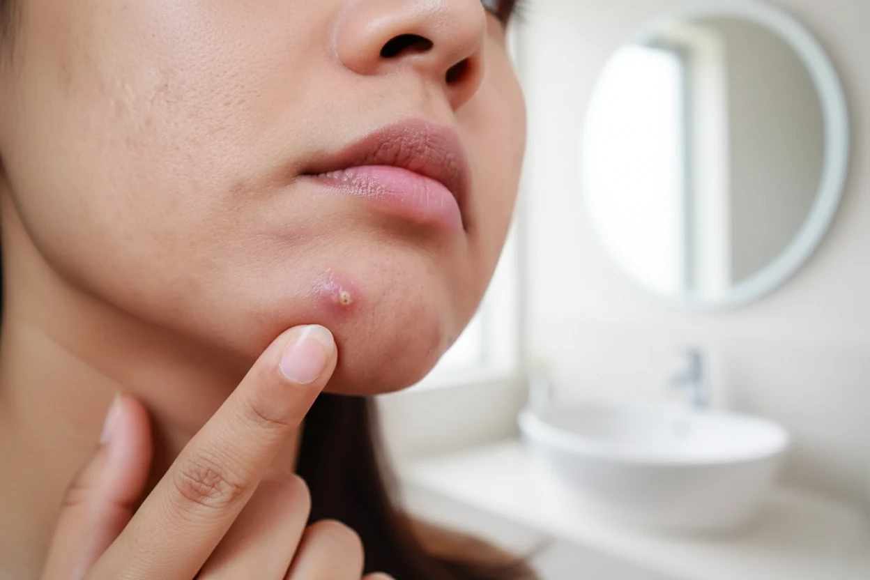
- Superficial hemangiomas: Bright red, raised, and rubbery in texture, often described as "strawberry marks"41.
- Deep hemangiomas: Bluish or skin-colored swelling beneath the skin surface8.
- Mixed hemangiomas: Combination of superficial and deep components3.
The proliferative phase usually peaks by 5 to 7 months of age, during which the lesion grows rapidly2. After this, the growth stabilizes before the slow involution phase begins4.
Symptoms may include:
- Visible red or purple lumps on the skin1.
- Swelling or bulging in affected areas9.
- Possible pain or tenderness if the lesion ulcerates or is located in a sensitive area8.
Causes and Risk Factors
The exact cause of hemangiomas is unknown, but they are believed to arise from abnormal proliferation of endothelial cells, which form the lining of blood vessels1011. Research suggests that hemangiomas originate from stem cells and involve dysregulation of angiogenic factors such as vascular endothelial growth factor (VEGF) and basic fibroblast growth factor (bFGF)123.
Risk Factors
Several factors increase the likelihood of developing infantile hemangiomas:
- Female sex: Females are about three to five times more likely to develop hemangiomas than males211.
- Prematurity: Premature infants have a higher risk25.
- Low birth weight: Babies with low birth weight are more prone to hemangiomas2.
- Multiple gestation: Twins and other multiples have increased risk2.
- Caucasian ethnicity: IH are more common in white infants115.
- Possible placental abnormalities and advanced maternal age: These have been suggested but are less well established2.
Most hemangiomas occur sporadically, with rare familial cases reported10.
Diagnosis and Testing
Hemangiomas are primarily diagnosed through clinical history and physical examination138. The typical appearance and timing of onset usually allow for straightforward diagnosis.
Additional diagnostic tools may be used in certain cases:
- Ultrasound: Useful for assessing deeper or atypical lesions and determining blood flow characteristics133.
- Magnetic Resonance Imaging (MRI): Provides detailed soft tissue imaging for complex or large hemangiomas, especially those involving internal structures13.
- Computed Tomography (CT): Occasionally used for deep or complicated lesions, though less common due to radiation exposure13.
- Biopsy: Rarely needed but may be performed if malignancy is suspected or diagnosis is uncertain13.
Histopathological examination reveals proliferating endothelial cells and vascular channels, confirming the diagnosis13.
Hemangioma Treatment Options
Most infantile hemangiomas do not require treatment as they regress spontaneously over time24. However, treatment is indicated when hemangiomas cause functional impairment, ulceration, bleeding, or risk of permanent disfigurement14.
Treatment options include:
- Oral propranolol: The first-line systemic therapy for problematic hemangiomas; it reduces growth by inhibiting blood vessel proliferation14115.
- Systemic corticosteroids: An alternative for hemangiomas not responsive to propranolol14.
- Topical beta-blockers (e.g., timolol): Used for smaller, superficial hemangiomas145.
- Laser therapy: Pulsed dye laser can treat residual blood vessels or ulcerated lesions14.
- Surgical excision: Reserved for lesions causing functional problems, disfigurement, or failure of medical therapy143.
Treatment choice depends on lesion size, location, and potential complications. Early intervention can minimize complications and improve cosmetic outcomes14.
“Hemangiomas, colloquially termed 'strawberry marks', are the most common benign tumor of infancy and are caused by endothelial cell proliferation.”
— Amal Chamli, Charles Nicolle Hospital11
Prevention Strategies
Currently, there are no known preventive measures for hemangiomas because the exact cause is unknown and risk factors such as prematurity and placental abnormalities are not modifiable2. Most infantile hemangiomas regress spontaneously and do not require treatment2.
Treatment is reserved for cases where the hemangioma causes functional impairment, ulceration, bleeding, or risk of permanent disfigurement14. Early recognition and specialist referral for high-risk cases can help manage complications effectively.
Potential Complications
While most hemangiomas resolve without problems, complications can occur depending on the size, number, and location of lesions:
- Ulceration: The most common complication, especially in areas where skin rubs or folds, such as lips or intertriginous zones148.
- Functional impairment: Hemangiomas near the eyes, airway, or mouth can interfere with vision, breathing, feeding, or speech15165.
- PHACE syndrome: Large or segmental facial hemangiomas may be associated with this syndrome, which includes brain, arterial, cardiac, and eye anomalies17.
- Hepatic hemangiomas: Can cause high-output cardiac failure or hypothyroidism in rare cases8.
- Airway involvement: May lead to respiratory distress requiring urgent treatment8.
Living With Hemangioma
Most children with hemangiomas live normal lives without lasting effects, as the lesions typically shrink and disappear over time4. However, some may have residual skin changes such as telangiectasias (small visible blood vessels), atrophy, or fibrofatty tissue6.
Large or complicated hemangiomas may require ongoing monitoring and multidisciplinary care. Imaging such as ultrasound or MRI can be used to evaluate internal or deep hemangiomas13.
Oral hemangiomas can interfere with feeding and speech, requiring specialized management15. Early treatment of problematic hemangiomas improves functional and cosmetic outcomes14.
Hemangioma FAQs
Q: When do most infantile hemangiomas disappear?
A: Approximately 50% of infantile hemangiomas resolve by age 5, and about 90% disappear by age 96.
Q: Are hemangiomas cancerous?
A: No, hemangiomas are benign (non-cancerous) tumors caused by abnormal blood vessel growth181.
Q: What treatments are available for hemangiomas?
A: Treatments include oral propranolol, corticosteroids, topical beta-blockers, laser therapy, and surgery, depending on the lesion's size, location, and complications143.
Q: Can hemangiomas cause complications?
A: Yes, complications such as ulceration, bleeding, functional impairment, and rare syndromes like PHACE can occur, especially with large or facial hemangiomas1714.