Dry eye disease (DED) affects a significant portion of the population worldwide, with prevalence estimates ranging from 5% to 34% depending on the population studied1. This multifactorial condition results from tear film instability or deficiency, leading to ocular discomfort, visual disturbances, and potential damage to the ocular surface2. Accurate diagnosis is essential to guide effective treatment and prevent complications such as vision impairment3. Diagnosis typically involves a combination of patient symptom questionnaires and a comprehensive eye examination using various clinical tests4.
Patient Symptom Questionnaires
The initial step in diagnosing dry eye disease involves assessing symptoms through validated questionnaires. The Ocular Surface Disease Index (OSDI) is widely used and consists of 12 questions that evaluate symptoms, functional limitations, and environmental triggers3. These questionnaires standardize patient-reported outcomes and correlate well with clinical findings5. They assess symptoms such as eye discomfort and visual disturbances, as well as functional impairments like difficulty reading or driving at night3. Environmental factors such as wind or dry air are also included to identify triggers3.
Patient history is equally important, encompassing symptom duration, severity, lifestyle factors, and family history6. This information helps guide the selection of further diagnostic tests during the eye examination6. Repeated use of questionnaires during follow-up visits allows clinicians to monitor treatment efficacy and disease progression7.
- The OSDI questionnaire evaluates dry eye symptoms and their impact on vision-related function7.
- Functional impairment related to dry eye, such as difficulty with reading or night driving, is assessed3.
- Environmental triggers like wind or dry air are included in symptom questionnaires3.
- Patient history including symptom duration and severity is essential8.
- Follow-up questionnaires help monitor treatment response7.
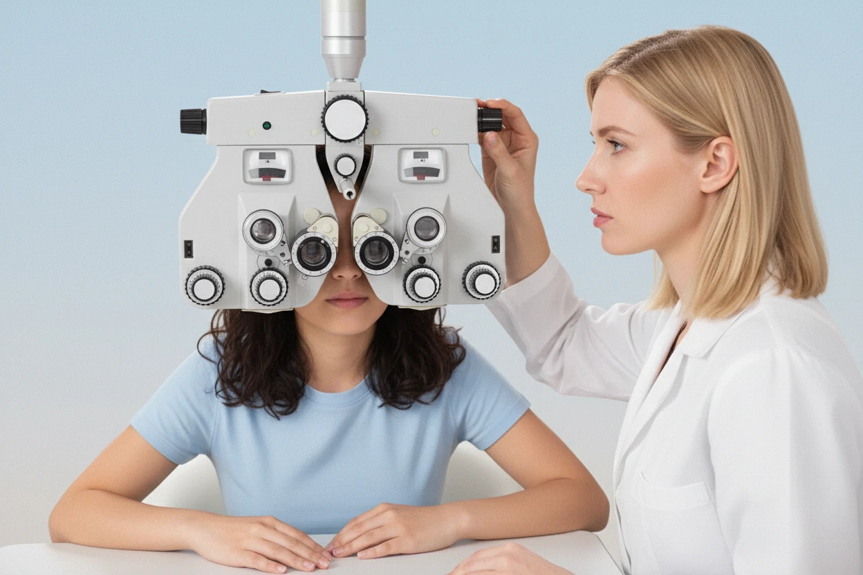
Comprehensive Eye Examination
Following symptom assessment, a comprehensive eye examination is performed to evaluate signs of dry eye disease9. This examination includes clinical evaluation of the ocular surface and tear film. Multiple diagnostic tests are used in combination to confirm the diagnosis, as no single test is sufficient9. Pupil dilation may be performed to facilitate detailed examination of the ocular surface and deeper ocular structures if needed9.
The comprehensive exam aims to detect ocular surface abnormalities, tear film instability, and tear production deficiencies. It provides objective data that complement patient-reported symptoms and guides tailored treatment plans4.
- Clinical examination is the gold standard for diagnosing dry eye syndrome10.
- Multiple diagnostic tests are combined to establish diagnosis9.
- Pupil dilation may be used to examine deeper ocular structures9.
- The exam evaluates signs such as redness, epithelial damage, and tear film quality4.
- Comprehensive evaluation helps differentiate dry eye from other ocular surface diseases6.
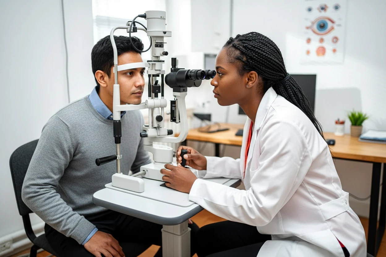
Slit Lamp Biomicroscopy Evaluation
Slit lamp biomicroscopy is a cornerstone of dry eye diagnosis, providing magnified visualization of the anterior eye structures including the cornea, conjunctiva, and eyelids9. This test allows clinicians to detect corneal epithelial defects, eyelid abnormalities, tear film instability, and blink dysfunction9. The slit lamp uses safe light intensity and is generally well tolerated by patients, who should report any discomfort during the exam9.
While slit lamp findings support the diagnosis, they are not definitive alone and are usually combined with other tests such as Schirmer’s test and tear break-up time (TBUT)54. The examination can reveal signs of ocular surface inflammation and damage that are common in dry eye disease4.
- Slit lamp biomicroscopy provides detailed inspection of the ocular surface and tear film4.
- It detects corneal epithelial defects and eyelid inflammation9.
- The procedure is safe and patient comfort is monitored9.
- Slit lamp findings support but do not confirm dry eye diagnosis alone9.
- It is part of a battery of tests required for diagnosis9.
“All that’s needed to diagnose dry eyes are a history, a slit lamp, and a fluorescein strip.”
— Deepinder K. Dhaliwal, MD, LAc, University of Pittsburgh School of Medicine11
Schirmer's Tear Production Test
Schirmer’s test quantifies tear production by measuring the wetting length of filter paper strips placed in the lower eyelid for five minutes9. The test can be performed with or without topical anesthesia to reduce discomfort9. Patients typically close their eyes during the test, and the length of the moistened strip indicates tear volume9. A wetting length of less than 5 mm after five minutes suggests aqueous tear deficiency consistent with dry eye disease9.
An alternative to Schirmer’s test is the phenol red thread test, which uses a pH-sensitive thread to assess tear volume in a less invasive manner9. Although Schirmer’s test may cause mild irritation, it provides objective data that complement symptom assessment9.
- Schirmer’s test measures tear production using filter paper strips placed in the lower conjunctival sac9.
- The test duration is five minutes with eyes closed9.
- A wetting length less than 5 mm indicates aqueous tear deficiency9.
- Topical anesthesia may be used to reduce discomfort9.
- The phenol red thread test is a less invasive alternative9.
Tear Break-Up Time Assessment
The tear break-up time (TBUT) test evaluates tear film stability by measuring the time interval between a blink and the appearance of the first dry spot on the cornea after fluorescein dye instillation9. After the dye is applied, patients blink to distribute it evenly and then keep their eyes open without blinking while the clinician observes under cobalt blue light9. A TBUT of less than 5 seconds is indicative of tear film instability and dry eye disease9.
This test is simple, non-invasive, and widely used in clinical practice to assess the quality of the tear film54. TBUT helps identify tear film instability, a key factor in dry eye pathophysiology11.
- TBUT measures the interval between a blink and tear film breakup9.
- Fluorescein dye is instilled to visualize the tear film under blue light9.
- Patients blink to distribute dye evenly before measurement9.
- A TBUT less than 5 seconds indicates tear film instability9.
- The test is non-invasive and quick to perform4.
Additional Diagnostic Tests
Additional tests may be employed to support dry eye diagnosis and assess ocular surface damage and inflammation9. Fluorescein and lissamine green staining highlight corneal epithelial defects and devitalized cells, respectively, providing visualization of ocular surface damage common in dry eye disease9. Lissamine green is better tolerated than rose bengal and is used to stain the conjunctiva12.
Tear film osmolarity measurement quantifies tear salt concentration, with elevated osmolarity indicating tear film instability and dry eye severity9. Point-of-care devices can perform this test quickly in the clinical setting12. The matrix metalloproteinase-9 (MMP-9) test detects inflammation markers in tears, helping to identify inflammatory dry eye and guide anti-inflammatory treatment12.
- Fluorescein staining highlights corneal epithelial defects9.
- Lissamine green stains devitalized conjunctival cells and mucus9.
- Tear osmolarity testing measures salt concentration in tears9.
- Elevated tear osmolarity is a biomarker for dry eye severity9.
- MMP-9 testing detects ocular surface inflammation12.
“The treatment of dry eye has evolved from tear substitution alone to a rationally based therapeutic algorithm.”
— Elisabeth M. Messmer, Prof. Dr. med., Ludwig Maximilian University of Munich1
Screening for Underlying Conditions
Dry eye diagnosis can be complicated by symptom overlap with other ocular surface diseases and systemic conditions59. Common systemic diseases associated with dry eye include Sjögren’s syndrome, rheumatoid arthritis, lupus-diagnosis-process-and-testing-methodslupus-diagnosis-process-and-testing-methodslupus-butterfly-rash-symptoms-causes-and-treatmentlupus-butterfly-rash-symptoms-causes-and-treatmentlupus erythematosus, diabetes mellitus, and scleroderma9. Eye care specialists often screen for these conditions through history, examination, and laboratory testing68.
Referral to specialists is appropriate when systemic diseases are suspected to ensure comprehensive management8. Early identification of underlying autoimmune or inflammatory diseases is crucial as they may require targeted treatment beyond dry eye therapies1.
- Dry eye symptoms overlap with allergic conjunctivitis and blepharitis9.
- Systemic diseases commonly linked to dry eye include Sjögren’s syndrome and rheumatoid arthritis9.
- Screening includes detailed history and laboratory tests8.
- Referral to specialists is recommended when systemic disease is suspected8.
- Early diagnosis of systemic conditions improves overall management1.
Diagnosis Summary and Review
Dry eye disease is a multifactorial condition characterized by tear film instability or deficiency, leading to ocular discomfort, visual impairment, and ocular surface damage2. Diagnosis requires a combination of patient symptom questionnaires and clinical tests including slit lamp biomicroscopy, Schirmer’s test, and tear break-up time4. Additional tests such as ocular surface staining, tear osmolarity, and inflammation markers improve diagnostic accuracy9.
“Dry eye disease is a multifactorial disease characterized by a persistently unstable and/or deficient tear film causing discomfort and/or visual impairment, accompanied by variable degrees of ocular surface epitheliopathy, inflammation, and neurosensory abnormalities.”
— Stephen Pflugfelder, MD, Baylor College of Medicine11
Clinical examination remains the gold standard for diagnosis, as no single test is definitive10. Early diagnosis and treatment are essential to prevent complications such as corneal scarring and vision loss3. Management should be tailored based on the underlying cause and severity of dry eye1.
| Diagnostic Test | Purpose | Key Diagnostic Criterion |
|---|---|---|
| Ocular Surface Disease Index (OSDI) | Symptom assessment | Patient-reported symptom severity |
| Slit Lamp Biomicroscopy | Ocular surface and tear film evaluation | Detection of epithelial defects and inflammation |
| Schirmer’s Test | Tear production measurement | Wetting length < 5 mm in 5 minutes |
| Tear Break-Up Time (TBUT) | Tear film stability assessment | Break-up time < 5 seconds |
| Fluorescein/Lissamine Green Staining | Ocular surface damage visualization | Presence of staining indicating epithelial damage |
| Tear Osmolarity | Tear film salt concentration | Elevated osmolarity indicates dry eye |
| MMP-9 Test | Inflammation marker detection | Positive test indicates ocular surface inflammation |
- Diagnosis combines symptom questionnaires and multiple clinical tests4.
- No single test confirms dry eye; a battery of tests improves accuracy9.
- Early diagnosis prevents complications such as vision impairment3.
- Tailored treatment depends on the type and severity of dry eye1.
- Regular monitoring with questionnaires and exams guides therapy7.