Breast cancer is the most common cancer among women worldwide and a leading cause of cancer mortality1. Early detection through screening mammography significantly reduces mortality by identifying cancers at earlier, more treatable stages2. Diagnosis involves a combination of imaging tests and tissue biopsy, followed by staging to determine the extent of disease and guide treatment decisions3. Understanding the diagnostic process and stages of breast cancer is essential for timely and effective care.
Mammograms for Breast Cancer Detection
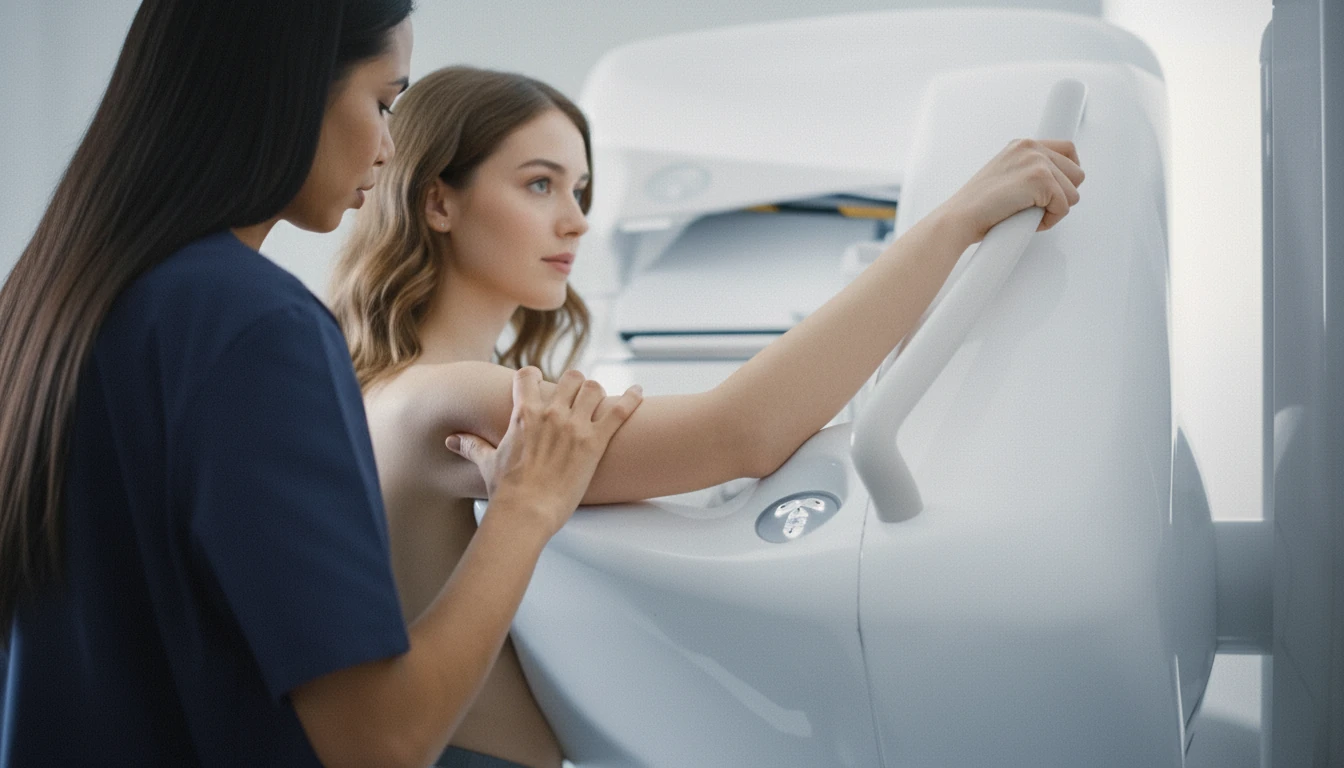
Mammography is a specialized X-ray technique that uses low-dose radiation to detect abnormalities in breast tissue, including masses and calcifications3. During the procedure, the breast is compressed between two plates to spread the tissue, which reduces overlap and improves image quality for better lesion detection34. While some discomfort is common during compression, severe pain is unusual4.
There are two main types of mammograms:
- Screening mammography is performed on asymptomatic women to detect breast cancer early before symptoms develop3.
- Diagnostic mammography involves additional views and is used when symptoms such as palpable lumps or nipple changes are present or when abnormalities are detected on screening mammograms34.
Mammography sensitivity decreases in women with dense breast tissue, where overlapping fibroglandular tissue can obscure lesions3. In such cases, adjunctive imaging like ultrasound or MRI may be recommended to improve cancer detection3.
Men have smaller breast tissue volumes, so mammography is less frequently used for breast cancer screening in men, except those at higher risk3.
“Women should consider 3D mammograms, especially if they have dense breasts. 3D screening mammography can increase the cancer detection rate in some cases, and studies suggest it can also reduce patient callbacks by up to 30%.”
— Cynthia A. Litwer, MD, Cedars-Sinai5
Breast Ultrasound Imaging
Breast ultrasound uses high-frequency sound waves to create images of breast tissue, helping to distinguish between cystic (fluid-filled) and solid masses3. A gel is applied to the skin, and a handheld transducer is moved over the breast to capture images4. Ultrasound is radiation-free and painless, though patients may feel mild pressure from the probe and a cold sensation from the gel34.
Ultrasound is particularly useful in the following scenarios:
- Characterizing lesions detected on mammography, especially in dense breasts34.
- Differentiating benign cysts from solid masses that may require biopsy3.
- Serving as an adjunct screening tool in women with dense breast tissue to improve cancer detection3.
Patients are encouraged to report any discomfort during the procedure to the technician3.
MRI Scans for Breast Evaluation
Magnetic resonance imaging (MRI) of the breast uses strong magnetic fields and radiofrequency pulses to generate detailed images of breast tissue3. Dedicated breast coils enhance signal reception and image quality. Intravenous administration of gadolinium-based contrast agents improves visualization of tumors and vascular abnormalities by highlighting areas of increased blood flow3.
Breast MRI is a non-invasive, painless procedure lasting 30 to 60 minutes34. However, some patients may experience anxiety or claustrophobia during the scan or discomfort related to the contrast injection3. Remaining still and breathing normally is important to avoid motion artifacts that can degrade image quality4.
MRI is indicated for:
- Further evaluation of suspicious clinical findings or inconclusive results from mammography or ultrasound3.
- Assessing tumor extent and multifocality in confirmed breast cancer cases to guide surgical planning3.
MRI is especially valuable for high-risk patients and those with dense breasts, as it can detect cancers not visible on mammograms3.
Breast Biopsy Procedures
A biopsy is the definitive test to confirm or rule out breast cancer after suspicious findings on imaging3. It involves removing a small sample of breast tissue or cells for microscopic examination by a pathologist3. Most biopsies reveal benign conditions, with the majority not showing malignancy3.
Biopsy methods include:
- Percutaneous needle biopsy, such as core needle biopsy or fine needle aspiration, guided by imaging techniques like mammography or ultrasound3.
- Surgical biopsy, which may be necessary if needle biopsy results are inconclusive or if the lesion is difficult to access3.
The choice of biopsy method depends on lesion characteristics, symptoms, and clinical judgment3. Post-biopsy pain varies based on the procedure type, tissue volume removed, and individual healing response. Pre-procedure counseling about sedation and pain control options is recommended to improve patient comfort3.
Breast Cancer Staging Explained
Staging is performed after diagnosis to determine the extent of cancer spread and to guide treatment decisions3. Breast cancer stages range from 0 to 4, with higher numbers indicating more advanced disease3.
The stages are defined as follows:
| Stage | Description | Tumor Size & Spread |
|---|---|---|
| 0 | Non-invasive cancer (ductal carcinoma in situ, DCIS) | Cancer cells confined to the ductal system3 |
| 1 | Early invasive cancer | Tumor ≤ 2 cm without lymph node involvement3 |
| 2 | Larger tumor or limited lymph node involvement | Tumor up to 5 cm, with or without limited nodes3 |
| 3 | Locally advanced cancer | Extensive lymph node involvement or chest wall invasion3 |
| 4 | Metastatic cancer | Spread to distant organs beyond breast and regional nodes3 |
Approximately 20% of breast cancers are detected at the DCIS stage, which is non-invasive and confined within the ducts3. Staging assessments may include physical exams, imaging tests such as bone scans, CT, MRI, or PET scans, and blood tests to evaluate organ function67.
Accurate staging helps predict prognosis and tailor treatment plans, including surgery, radiation, chemotherapy, and hormone therapy3.
Key Diagnostic Takeaways
“Studies show that routine breast cancer screening can reduce breast cancer deaths by one third and 40-50% in some trials.”
— Cynthia A. Litwer, MD, Cedars-Sinai5
- Breast cancer diagnosis relies on a combination of imaging tests—mammography, ultrasound, and MRI—and tissue biopsy for confirmation3.
- Mammography remains the primary screening tool and is recommended annually starting at age 40 for average-risk women38.
- Ultrasound is valuable for further characterizing lesions, especially in dense breasts, and is radiation-free and painless34.
- MRI provides high-resolution images and is useful for evaluating suspicious findings and assessing tumor extent, particularly in high-risk or dense breast patients3.
- Biopsy is essential to confirm cancer diagnosis and guide treatment, with multiple methods tailored to lesion characteristics3.
- Staging from 0 to 4 determines cancer spread and guides prognosis and therapy decisions3.