An echocardiogram is a common, noninvasive test that uses sound waves to create moving images of the heart. It helps healthcare professionals assess the heart’s structure and function, including how mcv blood test results meaning and normal range flows through the heart and its valves1. This test can diagnose a variety of heart conditions, including valve disease, heart failure, and congenital defects, and is often used to monitor treatment progress2.
Reasons for an Echocardiogram
Healthcare providers order echocardiograms for many clinical reasons. It is the primary non-invasive tool for evaluating the heart’s anatomy and function3. Common indications include:
- Evaluation of heart murmurs to determine their cause4.
- Assessment of symptoms such as chest pain, shortness of breath (dyspnea), palpitations, and fainting (syncope)4.
- Diagnosis and monitoring of myocardial infarction (heart attack) and heart failure4.
- Screening for heart disease in asymptomatic individuals, especially those with risk factors like hypertension5.
- Monitoring response to therapies for heart failure and valvular heart disease4.
- Assessment of myocardial ischemia (reduced blood flow to the heart) using stress echocardiography during exercise or pharmacologic stress4.
- Guiding therapeutic interventions such as volume resuscitation and vasoactive therapy in critically ill patients6.
In outpatient cardiology, hypertension accounts for about 23% of echocardiogram indications, while screening of asymptomatic patients accounts for 17%5. Echocardiography provides critical information that helps establish diagnoses and guides treatment decisions7.
An echocardiogram uses sound waves to show how blood flows through the heart and heart valves. The test can help a healthcare professional diagnose heart conditions.1
Echocardiogram Types
Echocardiography includes several modalities tailored to different clinical needs. The main types are:
- Transthoracic Echocardiogram (TTE): The most common type, performed by placing a transducer on the chest wall to obtain images of the heart from outside the body3. It is noninvasive and provides two-dimensional (2D) and three-dimensional (3D) images of heart structures and function83.
- Transesophageal Echocardiogram (TEE): Involves inserting an ultrasound probe into the esophagus to get higher-resolution images of the heart, especially the back structures and the aorta3. It is more invasive than TTE and used when detailed images are needed3.
- Stress Echocardiography: Combines echocardiography with exercise or pharmacologic stress to evaluate how the heart functions under stress and to detect myocardial ischemia3.
- Doppler Echocardiography: Measures blood flow velocity and direction through the heart and valves, useful for diagnosing valvular and congenital heart diseases8. It can be performed as part of TTE, TEE, or stress echo8.
- Three-Dimensional Echocardiography: Provides comprehensive 3D views of cardiac anatomy and function, often used for detailed valve assessment3.
Other specialized types include fetal echocardiography to assess the unborn baby's heart during pregnancy9.
“Transthoracic echocardiography (TTE) is routinely required during pre-participation screening in the presence of symptoms, family history of sudden cardiac death or cardiomyopathies in individuals under 40 years old, murmurs, abnormal ECG findings, or in the follow-up of athletes with a history of cardiovascular disease (CVD). TTE is a cost-effective first-line imaging modality to evaluate cardiac remodeling due to long-term, intense training (athlete's heart) and to rule out conditions at risk of sudden cardiac death, including cardiomyopathies, coronary artery anomalies, congenital, aortic, and heart valve diseases.”
— Elena Cavarretta, Italian Society of Sports Cardiology10
Preparation Steps
Preparation for an echocardiogram depends on the type of test being performed but is generally minimal:
- For TTE and Doppler echocardiography, no special preparation such as fasting is needed3.
- Patients should wear comfortable clothing and may need to remove clothing from the upper body to allow access for the transducer3.
- For stress echocardiography, patients should wear comfortable clothes suitable for exercise3.
- Patients must inform their healthcare provider about all medications, especially anticoagulants, as some medications may need to be withheld before the test3.
- For TEE, patients are typically instructed to fast for several hours before the procedure to reduce the risk of aspiration during sedation3.
- Patients should follow all instructions provided by their healthcare team to ensure a safe and accurate echocardiogram3.
💡 Did You Know? Echocardiography requires minimal preparation compared to other cardiac tests and does not involve radiation exposure113.
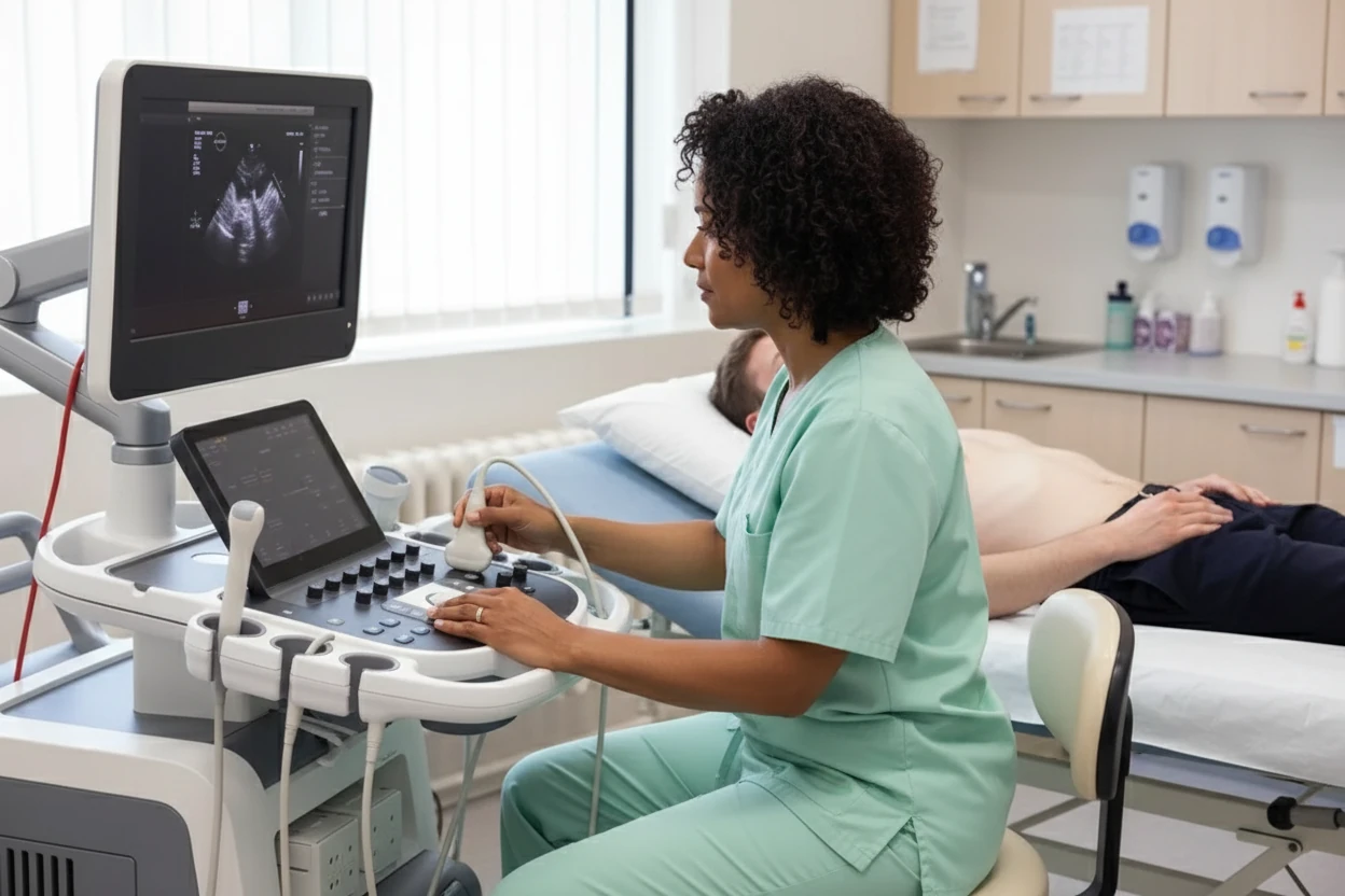
During the Echocardiogram Procedure
Echocardiography is usually performed in a hospital or outpatient clinic by a trained technician or physician and typically takes 30 to 60 minutes3. The procedure is painless and non-invasive, though some pressure may be felt from the transducer3.
During a Transthoracic Echocardiogram (TTE):
- The patient lies on a table, usually on their back or left side3.
- A gel is applied to the chest to improve sound wave transmission3.
- The transducer is placed on the chest and moved to different positions to obtain images of the heart3.
- Patients may be asked to hold their breath or change positions to improve image quality3.
- Doppler ultrasound is used to assess blood flow through the heart and valves during the test3.
During a Transesophageal Echocardiogram (TEE):
- The patient is sedated and given oxygen3.
- A thin, flexible probe with an ultrasound transducer is guided down the throat into the esophagus3.
- This allows for high-resolution images of the heart and aorta from inside the body3.
- The procedure may take up to 90 minutes12.
During a Stress Echocardiogram:
- Images are taken before and after exercise on a treadmill or stationary bike3.
- If the patient cannot exercise, medication may be given to simulate stress on the heart3.
- The test evaluates how the heart responds to physical activity or stress3.
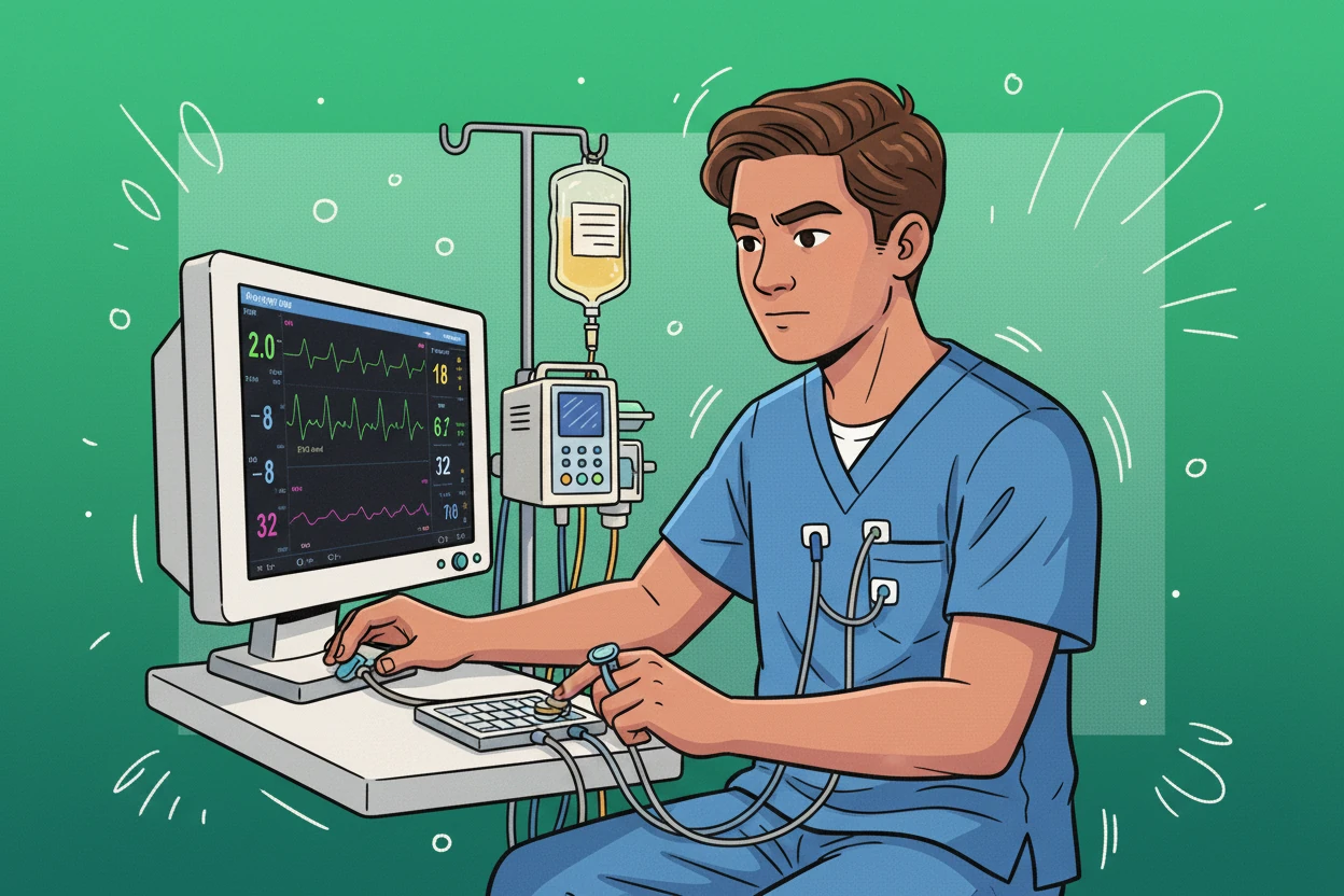
During an echocardiogram, sound waves bounce off the heart to create moving images that show heart structure and blood flow in real time.13
After the Echocardiogram Test
Most patients can resume normal activities immediately after an echocardiogram, as there is no recovery time needed for TTE or stress echocardiography113. After a TEE, some patients may experience mild throat discomfort or soreness, which usually resolves within a few hours3.
Patients should:
- Follow any specific post-test instructions from their healthcare provider3.
- Contact their provider if they experience unusual symptoms such as severe throat pain, difficulty swallowing, or chest discomfort after TEE3.
- Arrange for transportation home after TEE due to sedation effects2.
The images and data collected during the test are reviewed by a healthcare provider, who will discuss the results and any necessary next steps with the patient3.
Understanding Echocardiogram Results
Echocardiogram reports provide detailed information about the heart’s anatomy, function, and any abnormalities detected14. Key components of the report include:
- Measurements of cardiac chamber sizes and wall thickness14.
- Assessment of ventricular function, including ejection fraction (the percentage of blood pumped out with each heartbeat)14.
- Evaluation of heart valve structure and function, including detection of valve narrowing (stenosis) or leakage (regurgitation)14.
- Description of blood flow patterns through the heart using Doppler imaging14.
- Identification of structural defects, blood clots, or tumors within the heart14.
- Assessment of the pericardium (the heart’s outer lining) and major vessels14.
A normal echocardiogram indicates normal cardiac structure and function without significant pathology14. Abnormal findings may require further testing, treatment, or referral to a cardiologist3.
Echocardiograms can detect heart valve problems, heart muscle damage, congenital defects, and blood clots, helping guide diagnosis and treatment.9
Echocardiogram Risks and Safety
Echocardiography is generally safe and non-invasive, especially the transthoracic echocardiogram (TTE), which carries minimal risk3. However, some risks exist, particularly with more invasive types:
“Cardiac echocardiography is a safe and non-invasive test that provides the clinical team with important cardiac function data. This functional data is needed to guide diagnosis and treatment.”
— StatPearls, NIH15
- TTE risks: Mild discomfort from the pressure of the transducer or cold gel; no radiation exposure3.
- TEE risks: Invasive procedure with potential complications including esophageal injury, throat soreness, difficulty swallowing, minor bleeding, and adverse reactions to sedation3.
- Patients with pre-existing esophageal conditions are at higher risk for TEE complications3.
- Sedation used during TEE may cause side effects such as nausea or irregular heartbeats3.
- Stress echocardiography may cause temporary symptoms like chest pain, flushing, or irregular heartbeat; serious complications are rare2.
Patients should discuss any concerns or medical conditions with their healthcare provider before the test to minimize risks3.
| Echocardiogram Type | Risk Level | Common Risks |
|---|---|---|
| Transthoracic (TTE) | Minimal | Mild discomfort, gel coldness |
| Transesophageal (TEE) | Moderate (invasive) | Esophageal injury, sedation effects |
| Stress Echo | Low to moderate | Temporary chest pain, irregular heartbeat |
Echocardiogram Summary
Echocardiography is a versatile, noninvasive imaging technique that uses ultrasound to assess the heart’s structure and function. It is widely used to diagnose heart conditions, monitor disease progression, and guide treatment decisions113. The most common form, transthoracic echocardiography (TTE), is safe and painless, while transesophageal echocardiography (TEE) provides higher-resolution images but carries some risks due to its invasive nature3.
Preparation for echocardiography is generally minimal, with specific instructions depending on the type of test. The procedure is typically completed within an hour, and most patients can resume normal activities immediately afterward3. Echocardiogram results provide detailed insights into heart health, helping healthcare providers detect abnormalities and tailor care plans14.