Diastasis recti is a common condition affecting more than half of women after childbirth, characterized by the separation of the abdominal muscles along the midline12. This separation can cause a visible bulge in the abdomen and may persist for months or years postpartum, impacting physical function and quality of life13. Understanding the symptoms, causes, diagnosis, and treatment options is essential for effective management and recovery14.
Diastasis Recti Symptoms and Signs
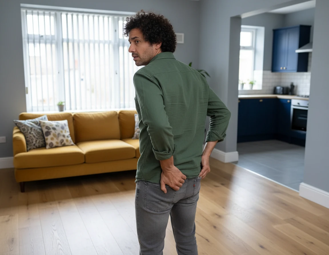
Diastasis recti abdominis (DRA) is marked by a separation of the rectus abdominis muscles along the linea alba, the connective tissue running down the midline of the abdomen56. A key visible symptom is a midline bulge or "pooch," often located between the sternum and the belly button, which becomes more prominent during muscle contraction73. Patients may notice coning or doming of the abdomen when engaging their abdominal muscles, such as during a crunch or sit-up movement73.
Other symptoms associated with diastasis recti include:
- Abdominal discomfort or a soft, jelly-like feeling around the belly button73
- Difficulty lifting objects or performing daily tasks due to core weakness1
- Pelvic floor dysfunction, including urinary incontinence and pelvic organ prolapse, where weakened pelvic muscles cause organs to descend lower in the pelvis73
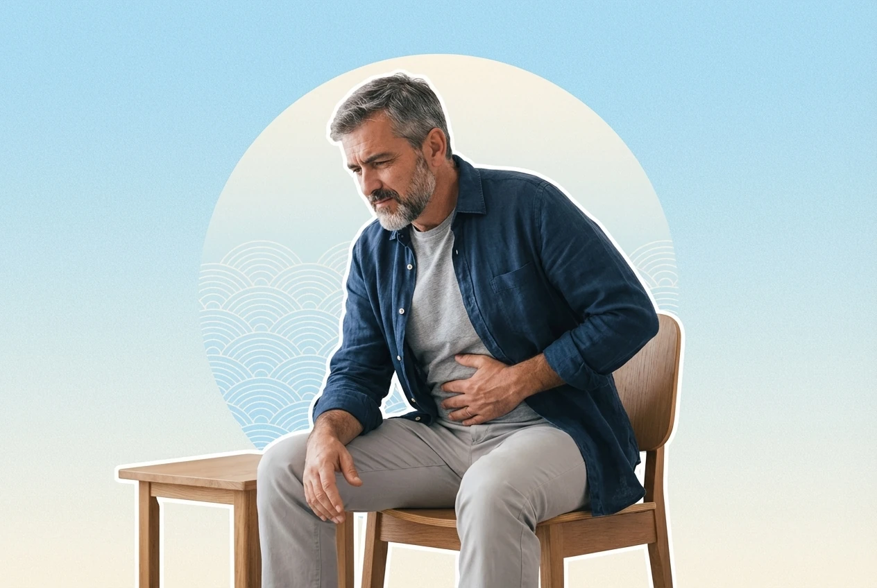
- Poor posture and lower back pain, which may range from dull aches to sharp pain, often worsened by lifting or bending73
While diastasis recti itself is usually not painful, the secondary effects such as musculoskeletal pain and pelvic floor issues can cause discomfort and functional impairment73.
Diastasis Recti Causes and Risk Factors
The rectus abdominis muscles run vertically along the front of the abdomen and are separated by the linea alba, a thin band of connective tissue73. During pregnancy-after-menopause-possibility-and-optionspregnancy-after-menopause-possibility-and-optionspregnancy, increased intra-abdominal pressure stretches this connective tissue, leading to muscle separation56. Hormonal changes in the second and third trimesters, including elevated levels of relaxin, estrogen, and progesterone, further loosen muscles and ligaments, weakening the linea alba76.
Additional risk factors for developing diastasis recti include:
- Multiparity (having multiple pregnancies) and cesarean delivery28
- Age between 30 and 39 years2
- Higher body mass index (BMI) and obesity, which increase abdominal pressure58
- Carrying multiples such as twins or triplets26
- Rapid weight gain during pregnancy58
- Having a large or heavy baby and closely spaced pregnancies1
These factors contribute to the mechanical and hormonal stresses that cause the linea alba to stretch and lose elasticity, preventing the abdominal muscles from returning to their normal position postpartum16.
Diagnosing Diastasis Recti
Diagnosis of diastasis recti typically involves a physical examination to assess the gap between the rectus abdominis muscles and to rule out other conditions such as hernias, which involve fascial defects56. A common clinical sign is a palpable or visible separation greater than two finger widths, often measured with the patient lying on their back with knees bent and shoulders slightly lifted15.
Ultrasound imaging is considered a reliable and accurate tool to measure the inter-recti distance (IRD)910. The European Hernia Society defines diastasis recti as a linea alba widening exceeding 2 cm611. Recent ultrasound diagnostic criteria specify:
- IRD greater than 2 mm at 3 cm below the umbilicus
- IRD greater than 20 mm at the umbilicus
- IRD greater than 14 mm at 3 cm above the umbilicus12
Measurement can be done at rest and during muscle activation, as the IRD may vary with posture and contraction12. If a separation is detected, consultation with a healthcare provider or physical therapist is recommended for further assessment and management56.
Diastasis Recti Treatment Options
Treatment for diastasis recti aims to restore core strength, improve symptoms, and reduce the abdominal gap56. Management depends on severity and associated symptoms and includes both conservative physical therapy and surgical options.
Conservative management is the first-line treatment and focuses on targeted core strengthening exercises to improve abdominal muscle function and support the linea alba1314. Exercises that engage the deep abdominal muscles, such as belly breathing, planks, glute bridges, and leg raises, may help narrow the separation and improve core stability1516.
Emerging therapies include SEMG (surface electromyography) biofeedback-assisted exercises combined with kinesiotaping, which have shown promise in enhancing muscle activation and reducing inter-recti distance1516. Physical therapists with expertise in postpartum care can tailor exercise programs to ensure safety and effectiveness, avoiding movements that may worsen the condition, such as traditional crunches or sit-ups15.
Key physical therapy considerations include:
- Avoiding exercises that cause abdominal doming or coning during contraction1
- Gradual progression to more challenging core exercises as muscle strength improves1
- Incorporating postural training and pelvic floor muscle rehabilitation73
- Educating patients on proper body mechanics to reduce intra-abdominal pressure during daily activities76
Surgical repair is considered for severe diastasis recti or when conservative treatment fails, especially if complicated by hernia formation1117. Surgical techniques range from open plication, where the separated muscles are sutured together, to minimally invasive laparoscopic approaches1819.
Surgical repair aims to:
- Close the abdominal gap and restore the integrity of the abdominal wall1819
- Improve core strength and posture2021
- Reduce symptoms such as back pain and functional impairment2021
Postoperative care includes wearing abdominal support for 6 to 8 weeks and avoiding strenuous activities or heavy lifting during recovery22. Long-term follow-up studies indicate sustained improvements in abdominal function and quality of life after surgery2021.
| Treatment Type | Description | Benefits | Considerations |
|---|---|---|---|
| Physical Therapy | Core strengthening exercises and kinesiotaping | Non-invasive, improves muscle function | Requires adherence and time |
| Surgical Repair | Open or laparoscopic muscle plication | Immediate closure of gap, symptom relief | Surgical risks, recovery period |
Preventing Diastasis Recti
Prevention strategies focus on modifiable risk factors to reduce the likelihood or severity of diastasis recti513. While some risk factors like carrying multiples cannot be changed, the following measures may help:
- Maintaining a healthy weight and avoiding excessive weight gain during pregnancy513
- Strengthening core muscles before and during pregnancy with safe exercises513
- Practicing good posture and deep breathing techniques to reduce abdominal strain76
- Avoiding excessive intra-abdominal pressure during physical activities, such as heavy lifting or straining76
- Using proper body mechanics, such as rolling onto the side when getting out of bed1
Evidence for specific preventive interventions remains limited, and individualized counseling is recommended to tailor strategies to each woman's needs613.
Conditions Related to Diastasis Recti
Diastasis recti is associated with several related conditions due to the weakening of the abdominal wall and core instability318. These include:
- Pelvic floor dysfunction, leading to urinary incontinence and pelvic organ prolapse318
- Increased risk of hernia development, where tissue protrudes through weakened fascia75
- Musculoskeletal pain, including lower back and hip pain, often exacerbated by movement or lifting73
- Functional impairments affecting posture, balance, and daily activities233
Addressing diastasis recti can help mitigate these complications and improve overall physical health313.
Living With Diastasis Recti
Living with diastasis recti involves managing both physical symptoms and psychological impacts3. Many postpartum women experience persistent abdominal bulging, discomfort, and functional limitations that affect daily life and self-image3. These challenges may lead to avoidance of activities such as lifting heavy objects or playing with children313.
“The abdominal muscles and surrounding tissue have to stretch to accommodate the growing fetus over the course of pregnancy.”
— Dr. Casey, Sports Medicine Physician24
A multidisciplinary approach is recommended, including:
- Physical therapy to improve core strength and function513
- Surgical options for severe cases or when conservative management is insufficient520
- Psychosocial support to address emotional concerns and improve quality of life313
Education about the condition and available treatments empowers patients to actively participate in their recovery and regain confidence in their bodies313.