Heart disease remains the leading cause of death worldwide, often presenting with silent or atypical symptoms that complicate early detection1. Timely diagnosis is essential to initiate effective treatment and improve patient outcomes1. A thorough evaluation combining medical history, physical examination, and a variety of diagnostic tests helps healthcare providers accurately identify heart attack symptoms in women conditions and guide management2.
Patient Medical History
The diagnosis of heart disease begins with a detailed medical history that focuses on symptoms and risk factors3. Key symptoms include chest pain, shortness of breath (dyspnea), and palpitations, which help clinicians assess the likelihood of cardiac disease3. Understanding the duration, severity, and variability of these symptoms is critical to determining urgency and guiding further testing3.
Risk factors such as family history of cardiovascular disease and previous cardiac events are important to establish during history taking4. Additionally, questions about current medications and recent infections or illnesses help clarify influences on heart symptoms and potential differential diagnoses3. This comprehensive history allows healthcare providers to stratify risk and tailor diagnostic pathways appropriately45.
Physical Examination
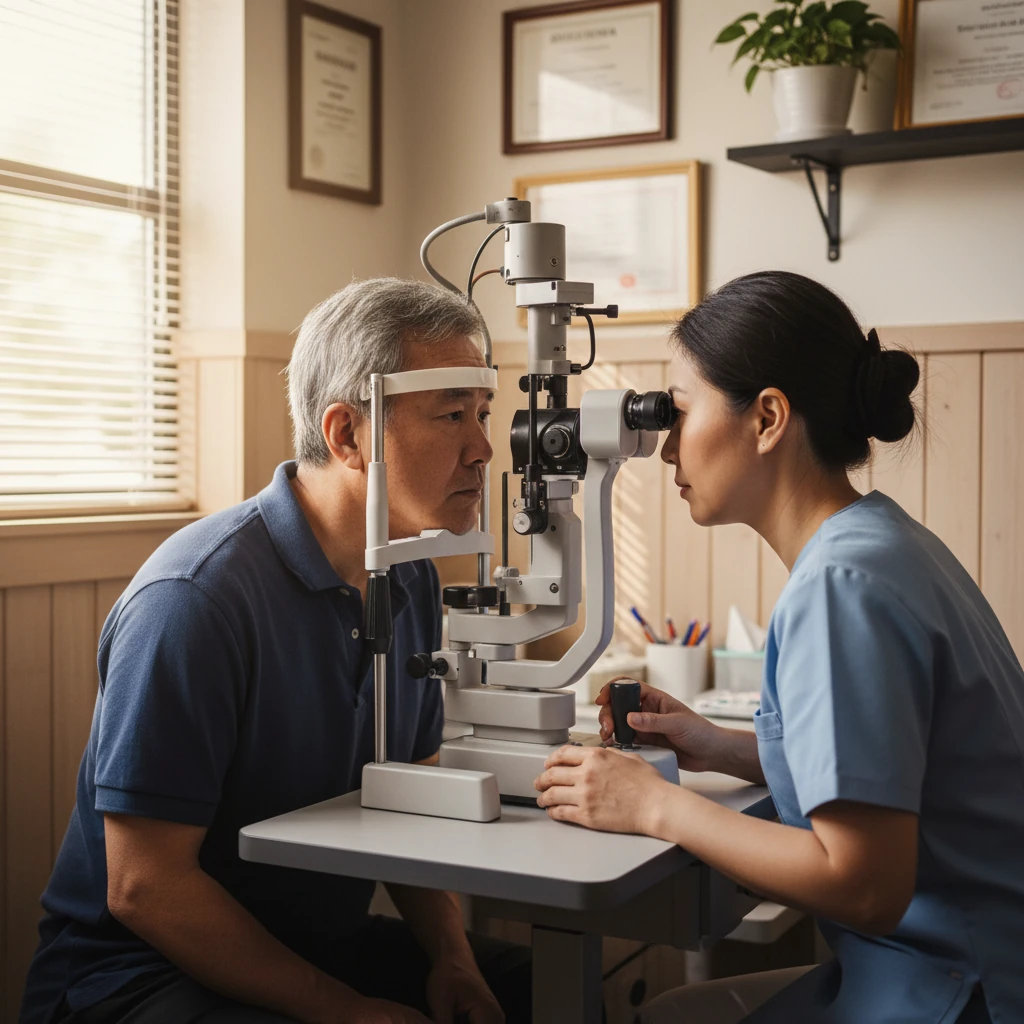
Physical examination is a cornerstone in diagnosing heart disease, providing direct clues about cardiac function and systemic involvement6. Clinicians assess vital signs, including blood pressure and pulse, which offer important information about heart rate, rhythm, and circulatory status3. Auscultation of the heart and lungs is performed to detect abnormal heart sounds such as murmurs, gallops, or crackles that may indicate valvular disease or heart failure3.
Other physical signs include peripheral edema (swelling), jugular venous distension, and lung crackles, which suggest fluid overload and heart failure7. These findings help prioritize further diagnostic testing and support cost-effective clinical decision-making8.
- Key physical exam components include:
- Measurement of blood pressure and pulse regularity3
- Auscultation for murmurs and abnormal breath sounds3
- Assessment for peripheral edema and jugular venous distension7
- Evaluation of lung sounds for crackles or congestion3
- Identification of signs of volume overload or heart failure7
Diagnostic Blood Tests
Blood tests are routinely used to detect heart problems and evaluate risk factors9. Cardiac biomarkers such as troponins are critical for diagnosing myocardial infarction (heart attack) by detecting heart muscle damage9. B-type natriuretic peptide (BNP) levels help identify heart failure by indicating cardiac stress and fluid overload9.
Lipid panels measure cholesterol and triglycerides to assess atherosclerotic risk, while metabolic panels evaluate kidney and liver function, which can influence heart disease management9. Inflammatory markers like high-sensitivity C-reactive protein (CRP) provide insight into arterial inflammation contributing to cardiovascular disease910.
- Common blood tests in heart disease diagnosis include:
- Troponin for myocardial injury9
- BNP for heart failure assessment9
- Lipid profile to evaluate cholesterol and triglycerides9
- Metabolic panels for organ function9
- CRP as a marker of inflammation910
Chest X-Ray Imaging
Chest X-rays are non-invasive imaging tests that assess the size and shape of the heart and evaluate pulmonary vasculature3. They are useful for detecting cardiomegaly (enlarged heart), pulmonary congestion, and pericardial effusion, which can indicate heart failure or other cardiopulmonary conditions63.
Interpretation of chest X-rays often involves collaboration between radiologists and cardiologists to integrate findings with clinical and laboratory data3. This imaging modality provides rapid and accessible information that complements other diagnostic tests3.
Electrocardiogram (ECG/EKG)
The electrocardiogram (ECG or EKG) is a fundamental, quick, and painless test that records the heart's electrical activity9. It provides information on heart rate, rhythm, and conduction, helping detect arrhythmias, ischemia, and prior myocardial injury9. ECG changes can indicate acute myocardial infarction or structural abnormalities9.
Ambulatory ECG monitoring devices, such as the Holter monitor or the Zio patch, extend monitoring over 24 hours or up to 14 days, respectively, improving detection of intermittent arrhythmias not captured on standard ECGs1112.
- ECG features and uses include:
- Recording cardiac electrical impulses and rhythm9
- Detecting arrhythmias and ischemic changes9
- Portable use in various clinical settings9
- Extended monitoring with Holter or Zio patch for intermittent arrhythmias1112
“Patients presenting with signs or symptoms of acute coronary syndrome should undergo timely electrocardiographic acquisition and interpretation, as ECG is central to the diagnosis of myocardial infarction.”
— Jacinda E. Tamis-Holland, American Heart Association13
Echocardiogram Ultrasound
Echocardiography uses ultrasound waves to create real-time images of the heart's structure and function14. It evaluates left ventricular ejection fraction, which measures the heart's pumping efficiency, and assesses valve morphology and function14. Echocardiograms can detect valvular stenosis or regurgitation, intracardiac masses such as thrombi or tumors, and cardiac muscle thickness and contractility14.
This modality is widely recommended for diagnosis, risk stratification, and ongoing monitoring of patients with known or suspected heart disease1415. Typical echocardiogram exams last between 30 to 60 minutes14.
- Echocardiogram capabilities include:
- Real-time imaging of cardiac chambers and valves14
- Measurement of ejection fraction and contractility14
- Detection of valvular disease and intracardiac masses14
- Assessment of cardiac muscle thickness14
- Monitoring disease progression or response to treatment14
Cardiac CT Scan
Cardiac computed tomography (CT) provides high-resolution, three-dimensional images of coronary arteries and cardiac structures16. CT angiography evaluates coronary artery plaque, stenosis, and cardiac chamber morphology, aiding in the diagnosis of coronary artery disease and pericardial disease16.
CT imaging protocols are typically rapid, lasting less than 15 minutes, and can identify pericardial effusion, coronary calcifications, and myocardial fibrosis16. This non-invasive modality complements other diagnostic tests by providing detailed anatomical information16.
Heart MRI Scan
Cardiac magnetic resonance imaging (MRI) uses magnetic fields and radio waves to produce detailed images of the heart's structure and function17. It is considered the gold standard for myocardial tissue characterization, assessing fibrosis, viability, and complex structural heart disease17.
MRI also supports evaluation of cerebrovascular and peripheral vascular diseases related to cardiac conditions17. Safety screening for implanted metallic devices is essential before MRI to avoid complications17.
- Cardiac MRI advantages include:
- High-resolution imaging of myocardial tissue and cardiac morphology17
- Assessment of myocardial viability and fibrosis17
- Evaluation of complex cardiomyopathies and structural abnormalities17
- Screening for related vascular diseases17
- Requirement for safety screening of implants17
Cardiac Catheterization
Cardiac catheterization is the gold standard invasive procedure for diagnosing coronary artery disease and guiding revascularization strategies4. It involves inserting a catheter into a blood vessel, usually the radial or femoral artery, and guiding it to the heart4. Contrast dye is injected to visualize coronary arteries under X-ray fluoroscopy, revealing blockages or stenoses4.
Due to its invasive nature, catheterization is reserved for patients with high suspicion or confirmed disease and provides direct anatomical and hemodynamic information414.
Cardiac Stress Test
Stress testing evaluates myocardial ischemia by increasing cardiac workload through exercise or pharmacologic agents16. Exercise treadmill testing is the most common form, assessing how the heart responds to physical activity and whether symptoms occur during exertion16.
For patients unable to exercise, pharmacologic stress agents like dobutamine or adenosine simulate the effects of exercise on the heart16. Vital signs are monitored during and after testing to ensure patient safety16.
- Stress test key points:
- Uses exercise or medication to increase cardiac workload16
- Detects ischemia and evaluates functional capacity16
- Exercise treadmill is the standard method16
- Pharmacologic agents used when exercise is not feasible16
- Continuous monitoring of heart rate, rhythm, and blood pressure16
Genetic Testing for Heart Conditions
Genetic testing complements clinical evaluation by identifying inherited cardiac disorders, enabling targeted screening and preventive strategies in at-risk individuals17. Family history of premature cardiovascular disease is a critical factor in risk stratification and may prompt early genetic evaluation4.
Inherited cardiomyopathies and arrhythmia syndromes can be detected through genetic testing, guiding personalized prevention and management plans17.
Related Condition Screening
Screening for related conditions is important because many diseases such as hypertension, diabetes, and valvular heart disease can contribute to or worsen heart failure and other cardiac conditions18. Comprehensive assessment helps identify comorbidities that affect diagnosis and treatment.
Regular screening includes blood pressure measurement, lipid and glucose testing, and evaluation for systemic diseases that impact cardiovascular health418.
Diagnosis Summary
Contemporary diagnosis and management of patients with myocardial infarction in the absence of obstructive coronary artery disease emphasize the importance of history, ECG, and cardiac troponin as central diagnostic tools13.
Diagnosing heart disease requires a multifaceted approach combining patient history, physical examination, laboratory tests, and imaging studies43. Symptoms like chest pain and dyspnea may overlap with non-cardiac conditions, necessitating careful differential diagnosis3. Multimorbidity is common, and a comprehensive evaluation helps avoid misdiagnosis and guides appropriate management43.
Diagnostic testing is individualized based on clinical findings and risk factors, ranging from non-invasive tests such as ECG and echocardiography to invasive procedures like cardiac catheterization43. Early detection and treatment improve prognosis and quality of life for patients with heart disease1.
- Summary of diagnostic steps:
- Detailed medical history and symptom assessment3
- Physical examination focusing on cardiac signs7
- Blood tests for biomarkers and risk factors9
- Non-invasive imaging including chest X-ray, ECG, echocardiogram, CT, and MRI141617
- Invasive evaluation with cardiac catheterization when indicated4