Ovarian cancer is the deadliest gynecological cancer deathscancer deaths, with over 75% of cases diagnosed at an advanced stage due to vague and nonspecific symptoms1. Early detection is challenging because symptoms often overlap with other common conditions, and no reliable screening test exists for the general population1. Diagnosis typically involves a combination of medical history, physical examination, imaging, blood tests, and tissue biopsy to confirm the presence and extent of cancer213.
Comprehensive Medical History & Risk Factor Assessment for Ovarian Cancer
A thorough medical history is crucial in assessing the risk of ovarian cancer. Symptoms at presentation are often vague and nonspecific, such as bloating, pelvic discomfort, or urinary changes, which contribute to delayed diagnosis1. Hereditary factors play a significant role; mutations in the BRCA1 and BRCA2 genes are linked to up to 20% of ovarian cancer cases4. Multimodal risk models that combine patient history, imaging findings, and serum biomarkers have improved the accuracy of adnexal mass assessment1.
Key points in medical history assessment include:
- Documenting family history of ovarian, breast, or related cancers41.
- Noting symptom onset, duration, and severity1.
- Assessing personal risk factors such as age, genetic mutations, and reproductive history1.
- Using risk assessment tools that integrate clinical and laboratory data1.
Pelvic Examination: What to Expect During an Ovarian Cancer Check
The pelvic exam remains the primary screening tool for ovarian cancer despite advances in other diagnostic methods5. During this exam, a healthcare provider palpates the uterus, ovaries, and surrounding pelvic organs to detect abnormalities6. However, early-stage ovarian tumors are often too small to be felt, which limits the exam’s sensitivity5. Consequently, 60% to 70% of patients present with advanced disease at diagnosis5.
Pelvic examination involves:
- Visual inspection of external genitalia, vagina, and cervix6.
- Bimanual palpation to assess organ size, shape, and tenderness6.
- Checking for signs of ascites (fluid in the abdomen) or masses6.
Diagnostic Imaging for Ovarian Cancer: Ultrasound, CT, MRI & PET Scans
Imaging studies are essential for detecting ovarian abnormalities, assessing disease extent, and guiding treatment planning. The main imaging modalities include transvaginal ultrasound, computed tomography (CT) scan, and magnetic resonance imaging (MRI) scan27.
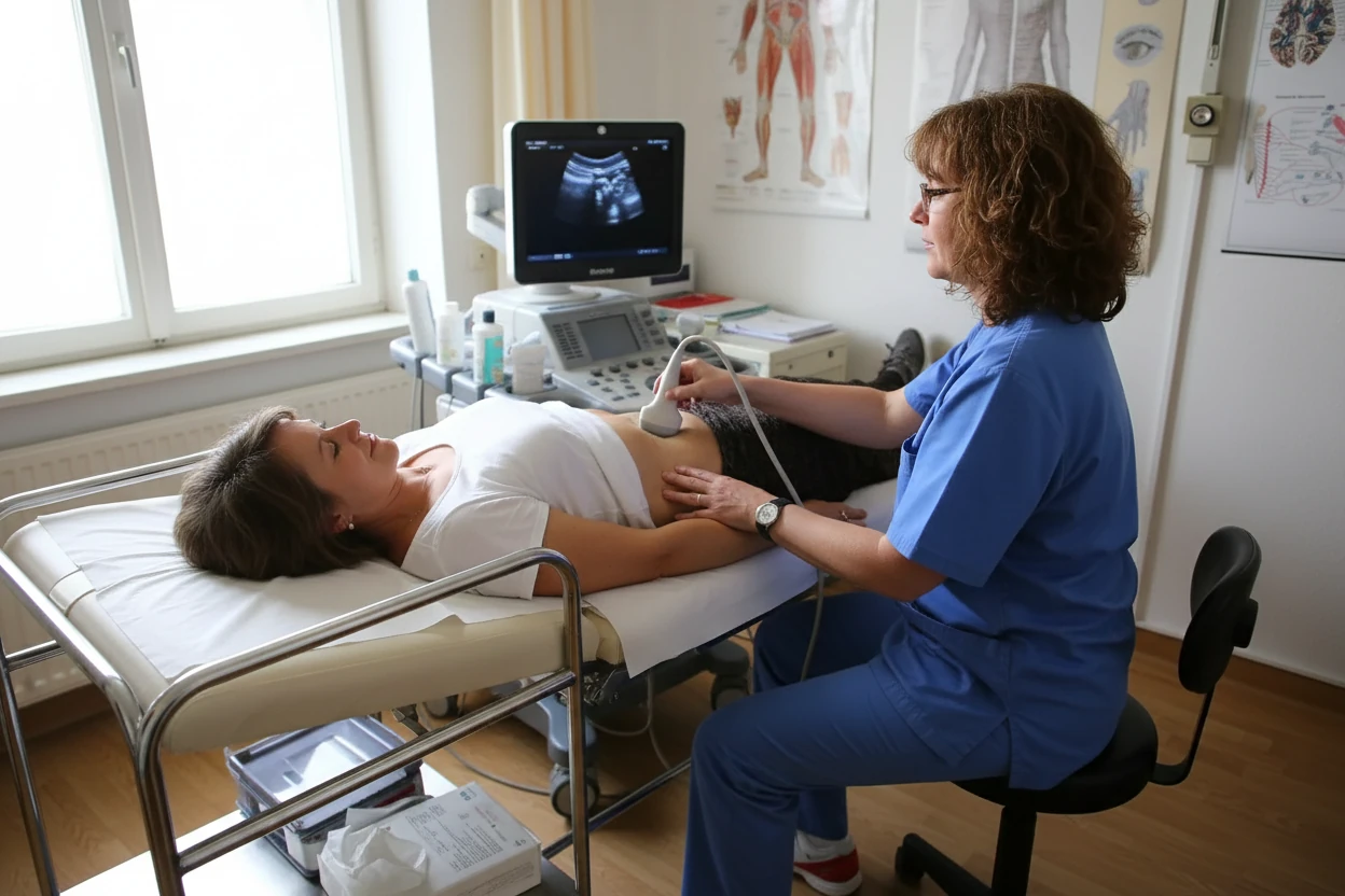
Transvaginal Ultrasound
Transvaginal ultrasound (TVS) is a sensitive tool for detecting ovarian abnormalities by inserting an ultrasound probe into the vagina to visualize the ovaries and uterus7. It accurately assesses ovarian volume and morphology, helping differentiate cystic from solid masses7. TVS is integral to ovarian cancer screening trials but has limited specificity for malignancy, as many benign masses can appear suspicious78.
Advantages of TVS:
- Non-invasive and widely available7.
- Provides detailed images of ovarian structure and blood flow7.
- Useful in initial evaluation of pelvic masses7.
Limitations:
- Cannot definitively distinguish benign from malignant tumors7.
- Operator-dependent accuracy7.
Computed Tomography (CT) Scan
CT scanning of the chest, abdomen, and pelvis is the imaging modality of choice for preoperative evaluation and follow-up in ovarian cancer patients29. CT helps map the disease extent, identify metastases, and evaluate lymph node involvement2. The radiological report should specify sites that may prevent optimal cytoreductive surgery, which is critical for treatment planning2.
Key roles of CT scan:
- Assessing tumor size and spread beyond the pelvis2.
- Guiding biopsy procedures when needed2.
- Monitoring response to therapy and detecting recurrence2.
Magnetic Resonance Imaging (MRI) Scan
MRI is an alternative imaging option used in specialized centers for ovarian cancer diagnosis and staging2. It provides high-contrast images and can be helpful when CT findings are inconclusive or when radiation exposure should be minimized2. MRI is particularly useful for evaluating soft tissue involvement and characterizing complex adnexal masses2.
MRI benefits:
- Superior soft tissue contrast2.
- Useful in patients with contraindications to CT contrast2.
- Helps in surgical planning by delineating tumor boundaries2.
Blood Tests for Ovarian Cancer: CA-125, HE4 & Other Biomarkers
Blood tests support ovarian cancer diagnosis by evaluating tumor markers and overall health status. They are also used to monitor treatment response and detect recurrence3.
CA-125 Blood Test
CA-125 is the most widely used tumor marker in ovarian cancer and is often considered the "gold standard" for monitoring disease3. Elevated CA-125 levels can indicate ovarian cancer but are not specific, as benign conditions like endometriosis, pelvic inflammatory disease, and pregnancy can also raise levels38. CA-125 is valuable for tracking chemotherapy response and detecting recurrence, with declining levels generally indicating treatment effectiveness3.
Important considerations for CA-125 testing:
- Not reliable as a screening test for asymptomatic women810.
- Useful in evaluating pelvic masses alongside imaging10.
- Elevated levels increase suspicion but do not confirm cancer3.
Other Tumor Marker Blood Tests
Additional tumor markers may complement CA-125, especially in specific ovarian cancer subtypes or younger patients311. These include:
- Human chorionic gonadotropin (HCG) and alpha-fetoprotein (AFP) for germ cell tumors11.
- HE4 (human epididymis protein 4), which may improve diagnostic accuracy when combined with CA-12512.
- Inhibin and lactate dehydrogenase (LDH) for sex cord-stromal tumors11.
- CEA and CA19-9 to evaluate for other malignancies or metastases11.
These markers help refine diagnosis and guide treatment decisions in selected cases1211.
Surgical Biopsy Procedure: Confirming Ovarian Cancer Diagnosis
A definitive diagnosis of ovarian cancer requires histopathological confirmation through tissue biopsy13. Biopsy is typically performed during surgery, either by removing the tumor or affected ovary13. No imaging or blood test can replace the need for tissue sampling to conclusively diagnose cancer13. In cases where surgery is not feasible, image-guided needle biopsy may be considered, although it carries a risk of spreading cancer cells13.
Paracentesis: Fluid Analysis for Ovarian Cancer Diagnosis & Spread
Ascites, or fluid accumulation in the abdomen, is common in advanced ovarian cancer. Paracentesis involves draining this fluid with a needle to relieve symptoms and obtain samples for cytological examination. Analysis of peritoneal fluid can detect cancer cells when surgical biopsy is not possible, aiding diagnosis.
Laparoscopy: Minimally Invasive Ovarian Cancer Diagnosis & Staging
Laparoscopy is a minimally invasive surgical procedure that allows direct visualization of the ovaries and surrounding tissues13. It enables surgeons to obtain tissue biopsies from suspicious lesions with less morbidity than open surgery13. Laparoscopy is useful for diagnosis, staging, and planning treatment in selected patients13.
Colonoscopy Evaluation: Assessing Bowel Involvement in Ovarian Cancer
While colonoscopy is primarily used to diagnose colorectal cancer, it may be indicated in ovarian cancer patients to assess for bowel involvement or metastasis. This evaluation helps determine the extent of disease spread and informs surgical planning.
Genetic Testing Options: BRCA1, BRCA2 & Hereditary Ovarian Cancer Risk
Germline genetic testing is increasingly important for ovarian cancer prevention and treatment4. Universal testing after diagnosis is recommended to identify inherited mutations, especially in BRCA1 and BRCA2 genes, which significantly increase ovarian cancer risk4. Other genes associated with hereditary ovarian cancer include those linked to Lynch syndrome4.
Benefits of genetic testing:
- Identifies patients who may benefit from targeted therapies such as PARP inhibitors4.
- Provides risk information for family members who may also carry mutations4.
- Guides personalized treatment and surveillance strategies4.
Ovarian Cancer Staging: FIGO System & Treatment Implications
Staging determines the extent of ovarian cancer spread and is critical for treatment planning and prognosis2. The FIGO system classifies ovarian cancer into four stages:
| Stage | Description | Key Features |
|---|---|---|
| I | Cancer confined to one or both ovaries | Tumor limited to ovaries or fallopian tubes |
| II | Cancer spread to pelvic organs | Involvement of uterus, fallopian tubes, or other pelvic tissues |
| III | Cancer spread beyond pelvis to abdomen or lymph nodes | Metastases to abdominal surfaces or lymph nodes |
| IV | Distant metastasis beyond abdomen and pelvis | Spread to liver, lungs, or distant organs |
| Sources:2 | ||
Screening for Related Conditions & Differential Diagnoses of Ovarian Masses
Screening for related conditions is important in ovarian cancer management. Genetic syndromes such as Lynch syndrome increase the risk of ovarian and other cancers, necessitating surveillance for colorectal and endometrial cancers4. Additionally, screening for benign gynecologic conditions that can mimic ovarian cancer symptoms, like endometriosis or pelvic inflammatory disease, helps avoid misdiagnosis3.
Ovarian Cancer Diagnosis Summary: Next Steps & Treatment Planning
Ovarian cancer diagnosis integrates clinical history, pelvic examination, imaging studies, blood tests, and histopathological confirmation213. Early symptoms are often nonspecific, leading to diagnosis at advanced stages in most patients1. Multimodal risk assessment combining these diagnostic tools improves accuracy and guides personalized treatment planning12.
Key diagnostic steps include:
- Detailed medical and family history assessment1.
- Pelvic examination to detect masses or ascites5.
- Imaging with transvaginal ultrasound, CT, and MRI to evaluate masses and disease spread27.
- Blood tests for CA-125 and other tumor markers312.
- Tissue biopsy via surgery or laparoscopy for definitive diagnosis13.
- Genetic testing to identify hereditary risk factors4.