Macular degeneration is a leading cause of vision loss in people over 50, affecting millions worldwide and resulting in irreversible damage to central vision1. This progressive eye disease primarily damages the macula, the central part of the retina responsible for sharp, straight-ahead vision2. While it rarely causes complete blindness, macular degeneration significantly impairs activities such as reading, recognizing faces, and driving3.
Understanding Macular Degeneration: Dry vs. Wet AMD Types
Macular degeneration is mainly classified into two types: dry (nonexudative) and wet (exudative or neovascular)4. Both types affect the macula but differ in their underlying mechanisms, progression, and severity of vision loss.
Dry Macular Degeneration
Dry macular degeneration accounts for approximately 85% to 90% of all cases and progresses slowly over time5. It is characterized by the gradual thinning and degeneration of the macula due to the accumulation of tiny yellow deposits called drusen beneath the retinal pigment epithelium (RPE)4. This buildup interferes with nutrient absorption, leading to atrophy of the RPE and loss of photoreceptor cells, which causes central vision to deteriorate4.
Dry AMD typically begins in one eye but can develop in both eyes over time, with patients having an increased risk of involvement of the fellow eye5. The disease progresses through three stages:
- Early: Presence of small drusen without vision loss6
- Intermediate: Larger drusen and pigment changes with mild vision symptoms6
- Advanced (geographic atrophy): Significant RPE and photoreceptor cell loss causing severe central vision impairment6
Vision loss in dry AMD is usually gradual, and peripheral vision remains intact7. However, dry AMD can convert to wet AMD, which is associated with more rapid and severe vision loss4.
Wet Macular Degeneration
Wet macular degeneration is less common, affecting about 10-15% of AMD patients, but it is more aggressive and causes rapid vision loss456. It occurs when abnormal mcv blood test results meaning and normal range vessels grow beneath the retina and macula, a process known as choroidal neovascularization5. These new vessels are fragile and prone to leaking blood and fluid, leading to hemorrhage, fluid accumulation, and scarring that damages the macula4.
Wet AMD often develops suddenly and can cause severe central vision loss if untreated5. It is considered an advanced form of AMD and may arise at any stage of dry AMD, especially in intermediate or advanced cases6. The leakage and scarring disrupt the normal architecture of the macula, resulting in distorted vision and blind spots4.
| Feature | Dry Macular Degeneration456 | Wet Macular Degeneration456 |
|---|---|---|
| Prevalence | 85-90% of AMD cases | 10-15% of AMD cases |
| Pathology | Drusen accumulation, RPE atrophy | Abnormal blood vessel growth, leakage |
| Progression | Slow, gradual vision loss | Rapid, severe vision loss |
| Vision Impact | Central vision loss, peripheral spared | Sudden central vision loss |
| Treatment Availability | Limited, mainly supportive | Anti-VEGF injections, photodynamic therapy |
| Sources:456 | ||
Macular Degeneration Symptoms: Early Signs & Vision Changes
The hallmark of macular degeneration is loss of central vision, which impairs the ability to see fine details directly ahead4. Common symptoms include:
- Difficulty reading or recognizing faces due to blurred central vision5
- Central blind spots (scotomas) appearing in the visual field5
- Distortion of straight lines, known as metamorphopsia, where lines appear wavy or bent4
- Increased need for brighter light when performing close-up tasks7
- Difficulty adapting to low light or night vision, although peripheral and night vision generally remain intact5
- Perception of glare or light distortion5
Despite these symptoms, macular degeneration rarely causes total blindness because peripheral vision is usually preserved4. Advanced disease can lead to legal blindness, defined as visual acuity of 20/200 or worse5.
Macular Degeneration Causes & Risk Factors: What Increases Your Risk?
Macular degeneration results from complex interactions between aging, genetic predisposition, and environmental influences that damage the macula8. The exact molecular mechanisms remain incompletely understood but involve oxidative stress, inflammation, and impaired blood flow to the retina4.
Risk Factors
Several factors increase the risk of developing AMD:
- Age: Most common in people over 50 years old4
- Family history and genetics: Several genes, including complement factor H polymorphisms, increase susceptibility94
- Race: More prevalent in white populations4
- Smoking: Significantly raises the risk and accelerates progression4
- Obesity: Associated with increased risk of progression from early to advanced AMD7
- Cardiovascular disease and hypertension: Linked to higher AMD risk due to vascular health impact4
- Diet: High intake of saturated fats increases risk, while diets rich in antioxidants and omega-3 fatty acids may be protective45
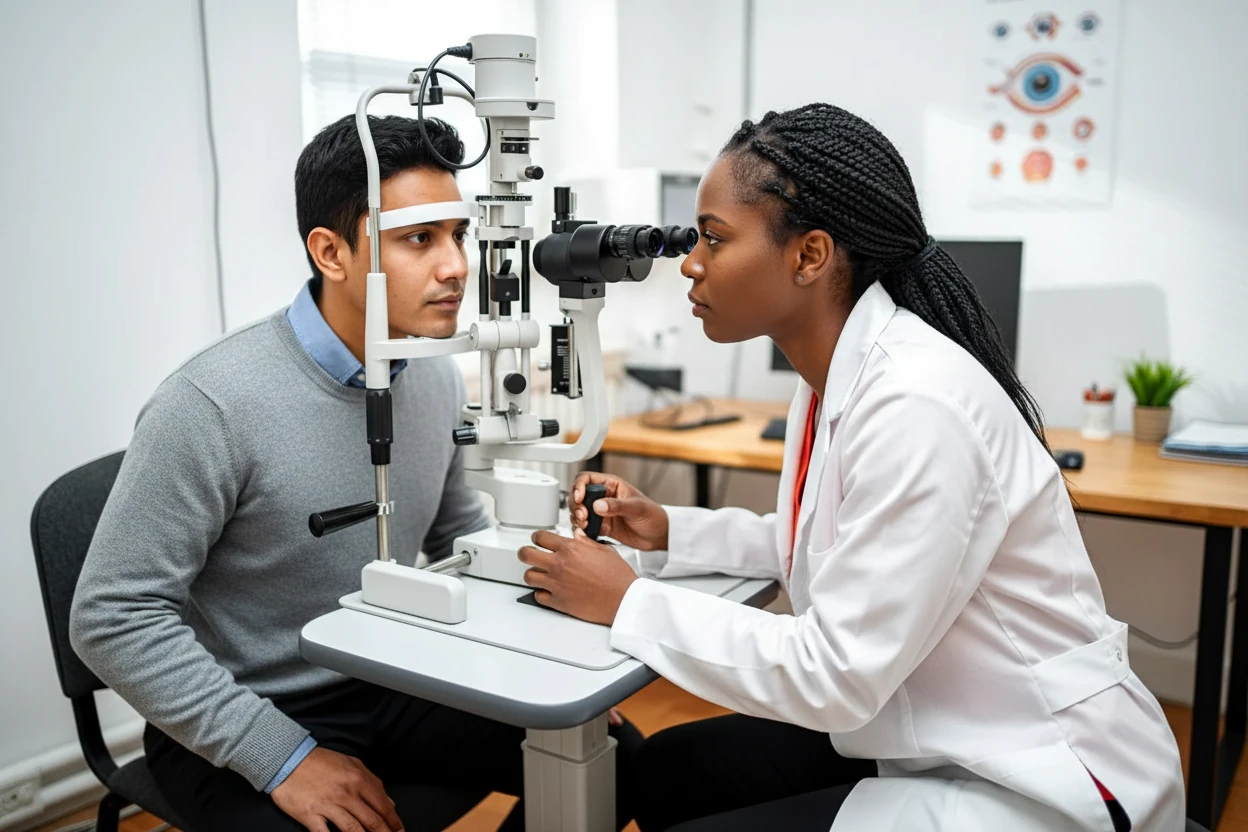
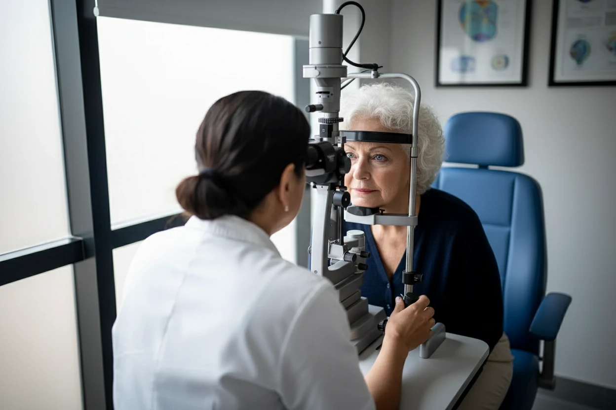
Diagnosing Macular Degeneration: Tests & Eye Exams
Diagnosis of AMD involves a comprehensive eye examination and specialized imaging to assess the retina and macula8. Key diagnostic tools include:
- Visual acuity testing to measure clarity of central vision4
- Dilated fundus examination to visualize the retina and detect drusen or abnormal blood vessels5
- Amsler grid test to detect visual distortions or blind spots in the central field10
- Optical coherence tomography (OCT) to obtain detailed cross-sectional images of retinal layers, revealing drusen, fluid, or atrophy4
- Fluorescein angiography involving intravenous dye injection to highlight leaking blood vessels in wet AMD5
- Optical coherence tomography angiography (OCTA) to visualize retinal and choroidal blood vessels noninvasively10
Stages of Macular Degeneration
Dry AMD is classified into stages based on drusen size and retinal pigment changes, which correlate with symptom severity4:
| Stage | Characteristics | Vision Impact |
|---|---|---|
| Early | Small drusen, no vision loss | Usually asymptomatic |
| Intermediate | Larger drusen, pigment abnormalities | Mild blurriness, difficulty in low light6 |
| Advanced | Geographic atrophy (RPE and photoreceptor loss) | Severe central vision loss |
| Sources:46 | ||
Wet AMD is considered an advanced stage due to the presence of neovascularization and rapid vision decline6.
Macular Degeneration Treatment Options: Managing AMD & Preserving Vision
Currently, there is no cure for macular degeneration, but treatments aim to slow progression and preserve remaining vision4. Early detection is critical for effective management8.
Supplements
For dry AMD, especially intermediate and advanced stages, nutritional supplements based on the Age-Related Eye Disease Study 2 (AREDS2) formulation may reduce the risk of progression5. These supplements include:
- Vitamin C (500 mg)11
- Vitamin E (400 IU)11
- Lutein (10 mg) and Zeaxanthin (2 mg)11
- Zinc (80 mg as zinc oxide)11
- Copper (2 mg as cupric oxide)11
Patients should consult healthcare providers before starting supplements, especially smokers, who should avoid beta-carotene due to increased lung cancer risk11.
“Faricimab will sometimes need to be administered frequently to keep the disease under control.”
— Carl Regillo13
Prescription Medications
Wet AMD is treated primarily with intravitreal injections of anti-vascular endothelial growth factor (anti-VEGF) drugs, which inhibit abnormal blood vessel growth and leakage345. Common anti-VEGF agents include:
- Aflibercept12
- Ranibizumab3
- Bevacizumab3
- Faricimab-svoa13
- Brolucizumab13
These injections are administered regularly to maintain disease control and may improve or stabilize vision13. Gene therapy is an emerging area with potential future benefits13.
Photodynamic Therapy
Photodynamic therapy (PDT) uses a light-activated drug to target and close abnormal blood vessels in wet AMD4. The treatment involves intravenous administration of a photosensitive drug followed by laser activation and eye protection5. PDT is less commonly used now but may be combined with anti-VEGF therapy in certain cases4.