Macular degeneration is a common eye condition affecting millions of people over the age of 50, making it a leading cause of irreversible vision loss in this age group1. This disease primarily damages the macula, the central part of the retina responsible for sharp, detailed vision needed for activities like reading and recognizing faces2. While early stages may show no symptoms, the condition can progress to cause significant central vision impairment, though peripheral vision usually remains intact3. Understanding the symptoms and signs across different stages of macular degeneration is crucial for early detection and timely management4.
Early Signs and Symptoms
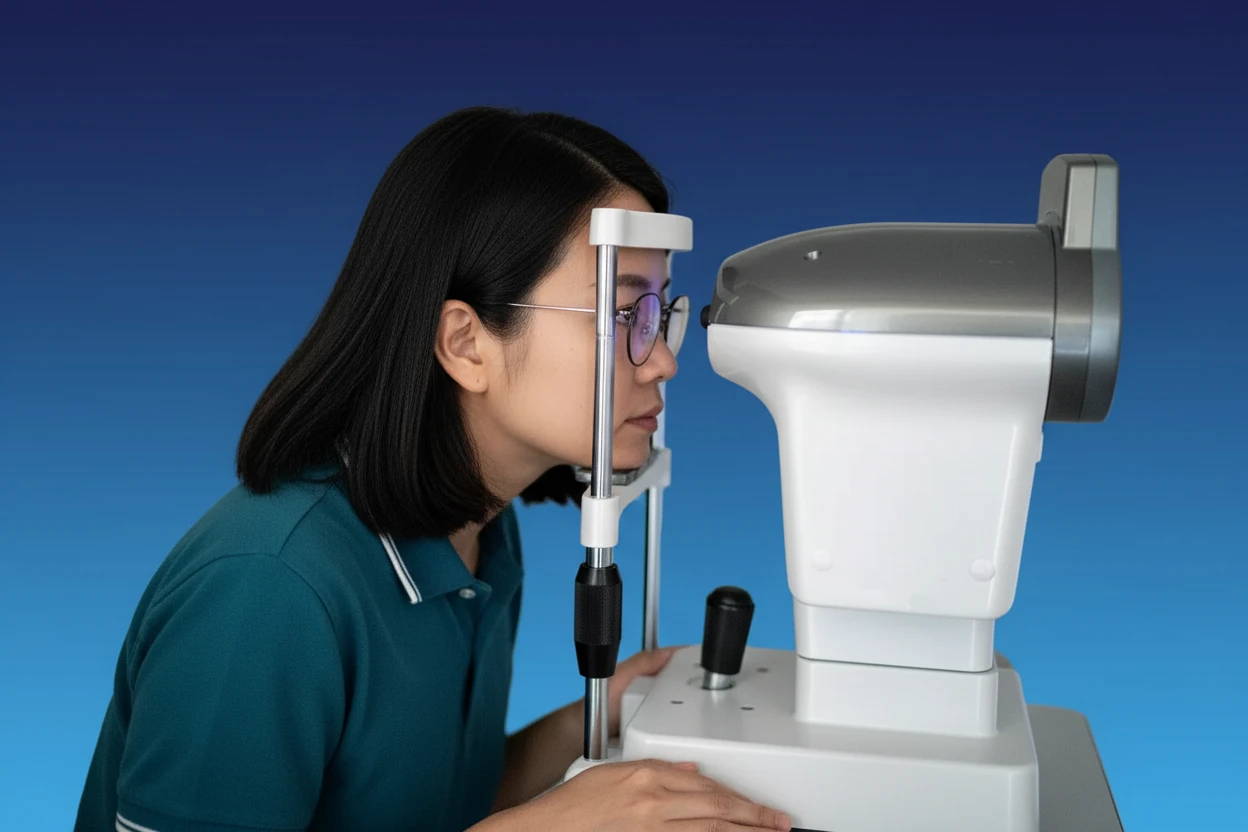
Early macular degeneration often develops without noticeable symptoms, making it difficult for individuals to detect on their own1. The hallmark of early age-related macular degeneration (AMD) is the presence of drusen—tiny yellow or white deposits under the retina—and pigmentary changes in the macula, which can only be identified through clinical examination or imaging techniques such as retinal photography or optical coherence tomography (OCT)256.
Drusen do not always lead to AMD but indicate a risk for disease development2. Early AMD may cause mild to moderate vision loss, but many patients remain asymptomatic during this stage5. Risk factors contributing to early AMD include increasing age, smoking, family history, and cardiovascular disease17.
Early detection through regular eye exams is essential because it allows for timely intervention to slow disease progression8. Clinical tools such as the Amsler grid test can help detect subtle visual distortions that may indicate early macular changes9.
💡 Did You Know?
Macular degeneration primarily affects central vision, meaning people with early AMD may not notice changes in their side (peripheral) vision3.
Intermediate Stage Symptoms
Intermediate AMD is characterized by larger drusen deposits and more pronounced pigmentary abnormalities beyond normal aging changes10. At this stage, some patients may begin to notice mild visual symptoms, although many remain asymptomatic10. Common complaints include blurred or distorted central vision, difficulty reading, and problems with recognizing faces211.
Visual distortions such as metamorphopsia—where straight lines appear wavy or bent—may develop during this stage122. Patients might also experience decreased contrast sensitivity, particularly affecting color perception2. Another early functional change under investigation is delayed rod-mediated dark adaptation (RMDA), which refers to slower adjustment to low light conditions and may be an early sign of AMD progression13.
As the disease advances within the intermediate stage, these symptoms can gradually worsen, signaling an increased risk of progression to late AMD, which includes geographic atrophy (advanced dry AMD) or neovascularization (wet AMD)10.
- Blurred or distorted vision2
- Metamorphopsia (distortion of straight lines)12
- Loss of contrast sensitivity, especially color-related2
- Difficulty adapting to low light13
- Mild visual impairment but often asymptomatic10
Advanced Macular Degeneration Signs
Advanced or late AMD is divided into two forms: geographic atrophy (dry AMD) and neovascular or exudative AMD (wet AMD)7. Both forms cause severe central vision loss, but wet AMD typically progresses more rapidly and can lead to blindness within a few years if untreated147.
Geographic atrophy involves the progressive loss of retinal pigment epithelium, choriocapillaris, and photoreceptor cells, leading to gradual but significant vision decline15. Wet AMD is marked by abnormal growth of new mcv blood test results meaning and normal range vessels under the retina (choroidal neovascularization), which leak fluid, lipids, and blood, causing swelling, scarring, and rapid vision loss15.
Symptoms of advanced AMD include:
- Severe central vision loss, often described as a dark or blind spot in the center of vision (central scotoma)127
- Visual distortion or metamorphopsia, where objects or lines appear bent or wavy12
- Loss of contrast sensitivity and color perception2
- Difficulty adjusting to low light or darkness2
- Possible visual hallucinations (Charles Bonnet syndrome) in cases of profound vision loss16
Peripheral vision is usually preserved, so total blindness is rare4. However, the loss of central vision significantly impairs daily activities such as reading, driving, and recognizing faces4.
| AMD Stage | Key Features | Progression Speed | Vision Impact |
|---|---|---|---|
| Early AMD | Small drusen, pigment changes, mild vision loss | Often asymptomatic | Minimal or no noticeable loss |
| Intermediate AMD | Large drusen, pigment abnormalities | Gradual | Mild to moderate visual symptoms |
| Advanced Dry AMD | Geographic atrophy, retinal cell loss | Slow | Severe central vision loss |
| Advanced Wet AMD | Neovascularization, leakage, scarring | Rapid (years) | Severe, often rapid vision loss |
| Sources:123 | |||
When to See a Doctor
Timely medical evaluation is critical, especially if symptoms suggest progression to wet AMD, which requires urgent treatment12. Patients should seek prompt ophthalmologic care if they notice:
- New or worsening visual distortions, such as straight lines appearing wavy or bent (metamorphopsia)12
- Sudden loss or blurring of central vision1
- Appearance of dark or blind spots in the center of vision (central scotoma)12
- Difficulty adjusting to low light or darkness2
- Any changes in color perception or contrast sensitivity2
Urgent referral to an ophthalmologist within one week is recommended if neovascular AMD is suspected to prevent irreversible vision loss12. The Rapid Access Referral Form (RARF) is used in some health systems to expedite evaluation and treatment17.
Regular eye exams are advised for individuals over 50 years, particularly those with risk factors such as smoking, family history, or cardiovascular disease1. Early diagnosis enables timely management, which can stabilize or slow disease progression8.
- Report any new central vision changes immediately1
- Attend regular eye exams, especially if over 50 or at risk1
- Seek urgent care if metamorphopsia or central scotoma develops12
- Follow recommended screening intervals based on age and risk9
- Use tools like the Amsler grid at home to monitor vision changes9
Urgent referral within one week to an ophthalmologist is necessary if neovascular AMD is suspected18.
Key Takeaways
- Macular degeneration primarily affects central vision by damaging the macula, the central part of the retina responsible for sharp, detailed vision23.
- Early AMD often has no symptoms but is marked by drusen and pigment changes detectable only by eye exams and imaging15.
- Intermediate AMD may cause mild visual symptoms such as blurred vision, metamorphopsia, and difficulty adapting to low light, but many patients remain asymptomatic1013.
- Advanced AMD leads to severe central vision loss; wet AMD progresses rapidly and can cause blindness within a few years if untreated714.
- Prompt medical evaluation and treatment, especially for wet AMD, are essential to preserve vision and slow disease progression128.
Macular Degeneration FAQs
What causes macular degeneration?
Macular degeneration is an age-related condition caused by damage to the macula, often influenced by genetic and environmental factors such as smoking, obesity, and cardiovascular disease167.
Can macular degeneration cause total blindness?
No, macular degeneration affects central vision but usually spares peripheral vision, so total blindness is rare43.
Is there a cure for macular degeneration?
Currently, there is no cure. Treatments like anti-VEGF injections can slow or stabilize wet AMD, but dry AMD has no approved cure and progresses slowly147.
How is macular degeneration diagnosed?
Diagnosis involves eye exams, visual acuity tests, Amsler grid testing, and imaging techniques such as OCT and fluorescein angiography to detect drusen, pigment changes, and abnormal blood vessels96.
What lifestyle changes can help reduce risk?
Quitting smoking, maintaining a healthy weight, managing cardiovascular health, eating a diet rich in fruits, vegetables, and omega-3 fatty acids, and regular exercise may reduce risk or slow progression161.