Lung cancer remains the leading cause of cancer-how-drinking-causes-breast cancer imaging costs deter follow up carecancer-effects-on-black-womencancer-related deaths worldwide, with early diagnosis significantly improving survival rates1. Accurate diagnosis involves a combination of clinical evaluation, imaging, laboratory testing, and functional assessments to guide treatment decisions2. This article outlines the comprehensive process and tests used to diagnose lung cancer effectively.
Initial Lung Cancer Assessment: Medical History & Physical Exam
The initial step in evaluating suspected lung cancer is a detailed medical history and physical examination3. This process helps identify risk factors and clinical features suggestive of lung cancer4. Physicians routinely assess respiratory symptoms such as cough, hemoptysis (coughing up blood), and dyspnea (shortness of breath)2. Lifestyle factors, particularly smoking history, and occupational exposures to carcinogens like asbestos and radon are also carefully evaluated2.
Physical examination may reveal signs indicative of lung cancer, including digital clubbing (enlargement of fingertips), lymphadenopathy (swollen lymph nodes), or respiratory distress2. Vital signs are assessed as part of the standard evaluation2. While history and physical exam alone cannot confirm lung cancer, they guide the selection of appropriate diagnostic tests2.
- Common symptoms prompting further evaluation include cough (50–75%), hemoptysis (25–50%), dyspnea (25%), and chest pain (20%)2.
- Occupational exposures to asbestos, radon, and secondhand smoke significantly increase lung cancer risk2.
- Physical signs such as clubbing and lymphadenopathy may suggest advanced disease2.
Blood Tests for Lung Cancer: Biomarkers & Tumor Markers
Blood tests are not diagnostic for lung cancer but are essential for assessing a patient’s overall health and organ function2. Routine blood work typically includes a complete blood count (CBC) and chemistry panels2. These tests help detect anemia, infection, and evaluate liver and kidney function, which may be affected by cancer or its treatment2.
Serum tumor markers such as CYFRA 21-1, carcinoembryonic antigen (CEA), and neuron-specific enolase (NSE) can be elevated in lung cancer but lack sufficient specificity for diagnosis5. Blood tests also play a role in monitoring treatment response and detecting disease progression or treatment side effects2.
- CBC helps identify anemia or infections that may complicate lung cancer management2.
- Blood chemistry tests assess liver enzymes and kidney function; abnormalities may indicate metastatic spread2.
- Serial blood tests are important for ongoing monitoring during treatment2.
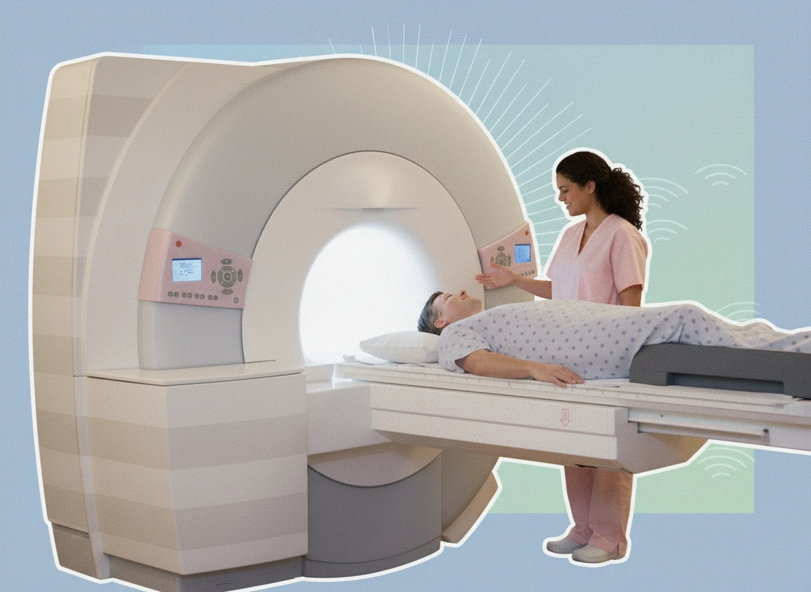
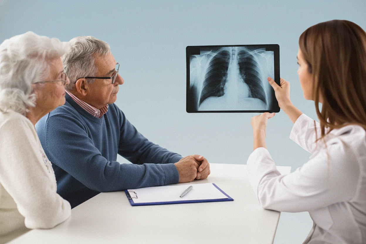
Lung Cancer Imaging: CT, PET Scans & X-rays for Diagnosis
Imaging is a cornerstone in lung cancer diagnosis, staging, and treatment monitoring6. The most common initial imaging test is a computed tomography (CT) scan, which provides detailed cross-sectional images of the lungs and chest7. CT scans are superior to chest X-rays in detecting lung nodules and assessing their characteristics8.
Positron emission tomography (PET) combined with CT (PET-CT) is recommended for staging to identify distant metastases by detecting metabolically active cancer cells7. Magnetic resonance imaging (MRI) is used selectively, particularly to evaluate brain metastases6.
- Chest X-ray is often the first imaging test but may miss up to 25% of lung cancers2.
- CT scans provide detailed visualization of lung lesions and guide biopsy procedures7.
- PET-CT scans detect cancer spread beyond the lungs, aiding in accurate staging7.
- MRI is valuable for assessing brain involvement in advanced disease6.
Biopsy & Laboratory Analysis: Confirming Lung Cancer Type
Tests for Diagnosis
Definitive diagnosis of lung cancer requires histopathological examination of tissue or cytology samples2. Several procedures are used to obtain these samples:
- Needle biopsy: A minimally invasive method where a needle, often guided by CT imaging, extracts tissue from lung nodules29.
- Bronchoscopy: A flexible tube with a camera is inserted through the mouth or nose into the airways to visualize and biopsy tumors210. Techniques such as endobronchial ultrasound (EBUS) enhance the ability to sample lymph nodes and peripheral lesions11.
- Sputum cytology: Examination of mucus coughed up from the lungs can detect cancer cells, especially in tumors originating in major airways29.
- Thoracentesis: Fluid is removed from around the lungs (pleural effusion) to check for malignant cells29.
- Mediastinoscopy: A surgical procedure to biopsy lymph nodes in the mediastinum (area between the lungs) for staging and diagnosis10.
Histopathological classification follows World Health Organization guidelines and increasingly incorporates molecular testing to identify genetic mutations that influence treatment212.
Tests for Cancer Spread
Determining the extent of cancer spread (staging) is essential for prognosis and treatment planning13. Imaging tests such as PET-CT, MRI, and bone scans are used to detect metastases69. Invasive procedures like mediastinoscopy or EBUS-guided needle aspiration help assess lymph node involvement11.
- Imaging identifies spread to lymph nodes, brain, bones, liver, and other organs13.
- Biopsy of lymph nodes confirms metastatic involvement2.
- Accurate staging guides surgical decisions and systemic therapy13.
Accurate tissue diagnosis and staging are critical to tailor lung cancer treatment effectively. Minimally invasive procedures like EBUS and needle biopsy reduce patient risk while providing essential diagnostic information102.
Pulmonary Function Tests (PFTs) Before Lung Cancer Treatment
Pulmonary function tests (PFTs) assess lung capacity and airflow to evaluate respiratory function before lung cancer surgery2. Common PFTs include spirometry, lung volume measurements, and diffusion capacity tests2. These tests help determine if a patient can tolerate lung resection and guide the extent of surgery2.
- Spirometry measures airflow and lung volumes2.
- Diffusion capacity assesses how well oxygen passes from lungs to blood2.
- PFT results influence surgical candidacy and postoperative management2.
Lung Cancer Staging: Determining Disease Extent & Treatment Options
Staging determines the size of the tumor, lymph node involvement, and presence of metastases to classify lung cancer and guide treatment13. Non-small cell lung cancer (NSCLC) uses the TNM system:
| Stage | Description | Tumor Size & Spread |
|---|---|---|
| 0 | Carcinoma in situ | Tumor confined to the innermost layer |
| I | Small tumor confined to lung | Tumor ≤ 3 cm, no lymph node involvement |
| II | Larger tumor or spread to nearby lymph nodes | Tumor > 3 cm or lymph node involvement |
| III | Locally advanced disease | Tumor invades nearby structures or multiple nodes |
| IV | Distant metastases | Cancer spread beyond chest to other organs |
| Sources:13 | ||
Small cell lung cancer (SCLC) is staged as limited (confined to one lung and nearby lymph nodes) or extensive (spread beyond one lung)13.
- Staging involves imaging, biopsy, and sometimes surgical exploration14.
- Accurate staging is crucial for prognosis and treatment selection13.
Screening for Related Conditions & Comorbidities in Lung Cancer
Early diagnosis of lung cancer is crucial for improving survival. Multidisciplinary teams and advanced diagnostic tools enable personalized treatment strategies12.
Lung cancer symptoms overlap with other respiratory diseases such as pneumonia, tuberculosis, and chronic obstructive pulmonary disease (COPD)2. Differential diagnosis is important to exclude these conditions and ensure appropriate management2.
Screening for lung cancer is recommended for high-risk individuals, typically adults aged 50–80 with a significant smoking history15. Low-dose CT scans are the preferred screening method and can detect early-stage cancers when treatment is more effective16.
- Screening reduces lung cancer mortality in high-risk populations2.
- Differential diagnosis requires careful evaluation to exclude infectious and inflammatory lung diseases2.
- Screening guidelines emphasize smoking history and age criteria15.
Lung Cancer Diagnosis Summary: Next Steps & Patient Review
Lung cancer diagnosis is a multi-step process beginning with clinical evaluation and risk assessment18. Imaging tests such as chest X-ray and CT scans identify suspicious lesions8. Definitive diagnosis requires tissue sampling through biopsy or cytology2. Staging using imaging and invasive procedures determines disease extent and guides treatment13.
Symptoms like cough, chest pain, and shortness of breath can mimic other respiratory conditions, making thorough evaluation essential2. Early diagnosis improves prognosis, with five-year survival rates exceeding 90% for stage IA disease but dropping below 10% for stage IV1.
- History and physical exam identify patients needing further testing18.
- Imaging and biopsy confirm diagnosis and stage disease2.
- Pulmonary function tests assess surgical candidacy2.
- Early detection and multidisciplinary care improve outcomes2.